Main menu
Common skin conditions

NEWS
Join DermNet PRO
Read more
Quick links

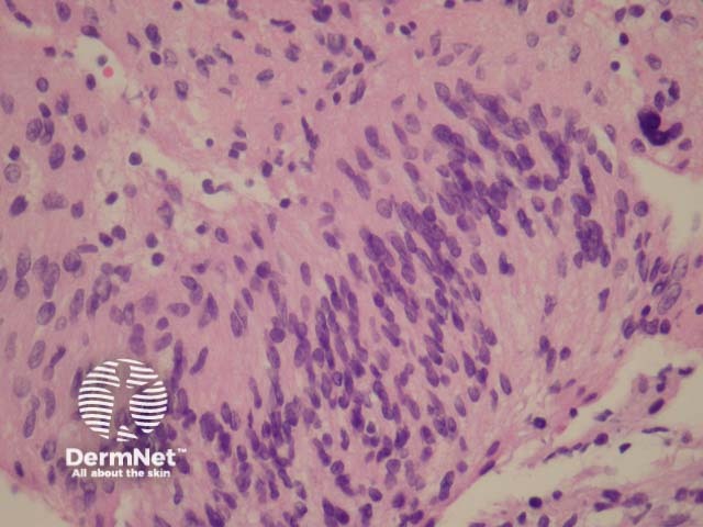
Figure 3
Keywords: Schwannoma, Histopathology-image, Pathology
In schwannoma, sections show an encapsulated well-circumscribed lesion beneath the uninterrupted epidermis. The tumour is composed of different areas composed of different cellular densities. More cellular areas (Antoni A, figure 1) are composed of a haphazard arrangement of bland cells with spindled and oval nuclei. Loose, less cellular areas (Antoni B, figure 2) are composed of a loose oedematous and mucinous stroma with fibrillar collagen. The vessels are prominent and often surrounded by a dense sclerosis.
© DermNet
You can use or share this image if you comply with our image licence. Please provide a link back to this page.
For a high resolution, unwatermarked copy contact us here. Fees apply.
Source: dermnetnz.org