Figure 3
Keywords: Merkel cell carcinoma, Histopathology-image, Pathology
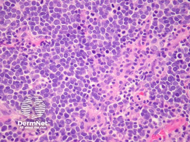
Merkel cell carcinoma is a neuroendocrine carcinoma composed of densely blue cells. The tumour is centered in the dermis with frequent involvement of the overlying epidermis (figures 1, 2) and may invade the subcutaneous fat. The tumour forms sheets, nests and rarely ribbons. The outlines of the cells often mold together or resemble lymphocytes (figures 3, 4). There are numerous mitoses, and there may be necrosis. Lymphovascular invasion is common.
© DermNet
You can use or share this image if you comply with our image licence. Please provide a link back to this page.
For a high resolution, unwatermarked copy contact us here. Fees apply.
Source: dermnetnz.org

