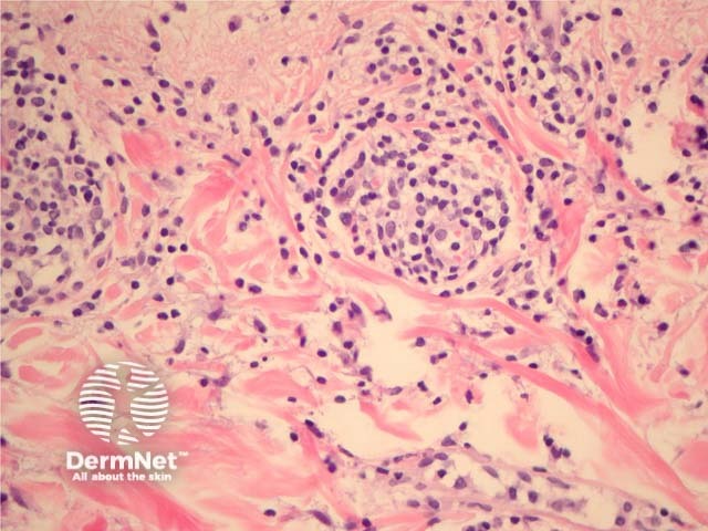
Figure 4
Keywords: Polymorphic light eruption, Histopathology-image, Pathology
In polymorphic light eruption, sections show a superficial and deep perivascular lymphocytic infiltrate (figure 1). There is often impressive papillary dermal oedema (figures 1, 2, 3). When the oedema is massive the lesions may resemble erythema multiforme clinically. The infiltrate is mainly lymphocytic but there may be intermixed eosinophils, neutrophils, and histiocytes (figure 4). The epidermal changes range from being almost normal to showing impressive spongiosis and acanthosis. There are often lymphocytes in the epidermis (exocytosis, figure 3). An interface dermatitis may be seen and associated apoptotic keratinocytes in the epidermis.
© DermNet
You can use or share this image if you comply with our image licence. Please provide a link back to this page.
For a high resolution, unwatermarked copy contact us here. Fees apply.
Source: dermnetnz.org

