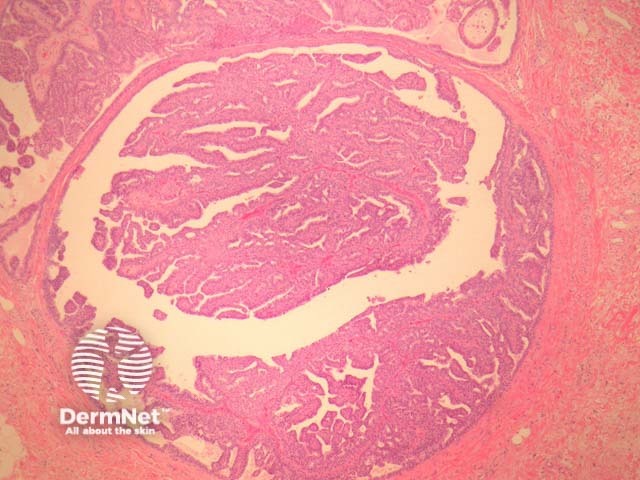
Figure 1
Keywords: Hidradenoma papilliferum, Histopathology-image, Pathology
Hidradenoma papilliferum is a well-circumscribed dermal nodule, usually without connection to the overlying epidermis (figure 1). Tubules and broad, elongated fronds form an arborizing pattern and are lined with a double-layered epithelium (figures 2, 3). The inner layer is comprised of cuboidal myoepithelial cells and the outer is tall columnar apocrine cells with a pale eosinophilic cytoplasm, which may demonstrate decapitation secretion. Larger fronds may have a fibrous core. Fibrous tissue surrounding the tumour may be compressed to form a pseudocapsule.
© DermNet
You can use or share this image if you comply with our image licence. Please provide a link back to this page.
For a high resolution, unwatermarked copy contact us here. Fees apply.
Source: dermnetnz.org

