Main menu
Common skin conditions

NEWS
Join DermNet PRO
Read more
Quick links

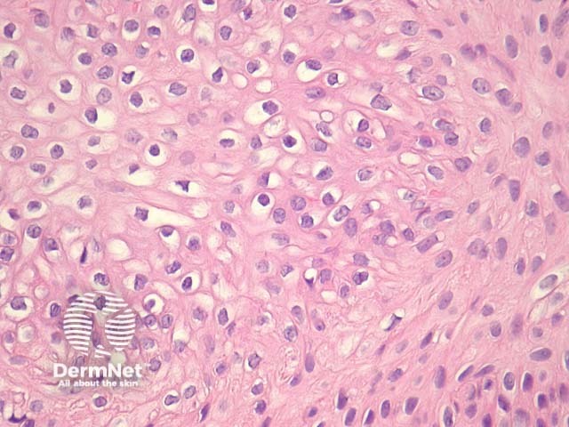
Figure 6
Keywords: Pilar sheath acanthoma, Histopathology-image, Pathology
In pilar sheath acanthoma, there is a lobular proliferation of benign squamous epithelium in the dermis (figure 1, 2). These lobules surround small cystic spaces. The lining cells may have a granular layer similar to an epidermoid cyst (figure 3) or have an attenuated granular layer similar to a trichilemmal cyst (figure 4).
© DermNet
You can use or share this image if you comply with our image licence. Please provide a link back to this page.
For a high resolution, unwatermarked copy contact us here. Fees apply.
Source: dermnetnz.org