Figure 4
Keywords: Prurigo pigmentosa, Histopathology-image, Pathology
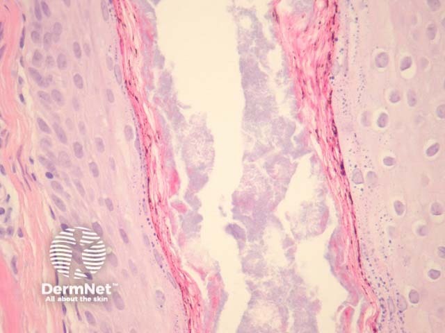
In well established prurigo pigmentosa, histopathologic examination reveals a sparse lichenoid infiltrate which also involves the superficial plexus and parakeratosis (figure 1). There are numerous dermal melanophages in later stages (figure 2). High power examination shows the lichenoid reaction is associated with apoptotic and necrotic keratinocytes present at all levels of the epidermis (figure 3). It is quite common to find a suppurative folliculitis and often hair follicles are filled with bacteria (figure 4).
© DermNet
You can use or share this image if you comply with our image licence. Please provide a link back to this page.
For a high resolution, unwatermarked copy contact us here. Fees apply.
Source: dermnetnz.org

