Main menu
Common skin conditions

NEWS
Join DermNet PRO
Read more
Quick links

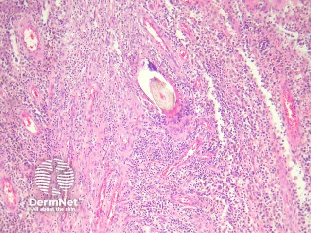
Figure 4
Keywords: Pilonidal sinus, Histopathology-image, Pathology
Sections show a dense inflammatory reaction usually occupying the entire dermis with erosion and ulceration of the overlying epidermis (figure 1). Free hair shafts are often seen coursing through the inflammatory focus (figure 2, arrow). Often, the free hair shafts are seen in clusters (figure 3). Dye used to outline the sinus tract for the surgeon may sometimes be seen. Surrounding the free hair shafts is a polymorphous infiltrate which may be rich in plasma cells and lymphocytes (figure 4). Foreign body-type giant cells and neutrophilic abscesses are also commonly observed.
© DermNet
You can use or share this image if you comply with our image licence. Please provide a link back to this page.
For a high resolution, unwatermarked copy contact us here. Fees apply.
Source: dermnetnz.org