Main menu
Common skin conditions

NEWS
Join DermNet PRO
Read more
Quick links

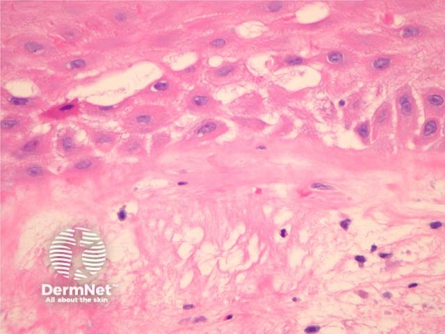
Figure 2
Keywords: Histopathology-image, Pathology, Radiation dermatitis
Chronic radiation dermatitis shows dermal sclerosis, elastosis and vascular ectasia overlying an epidermis which is often hyperkeratotic (figure 1). There may be epidermal spongiosis or impressive basal vacuolar change (figure 2). The dermal vessels are typically quite dilated in later stages (figure 3). Both the stroma fibroblasts and endothelial cells may show some hyperchromasia, enlargement and atypia (radiation fibroblasts, figure 3). There is often a mixed inflammatory response.
© DermNet
You can use or share this image if you comply with our image licence. Please provide a link back to this page.
For a high resolution, unwatermarked copy contact us here. Fees apply.
Source: dermnetnz.org