Basal cell carcinoma images
Go to basal cell carcinoma topic page
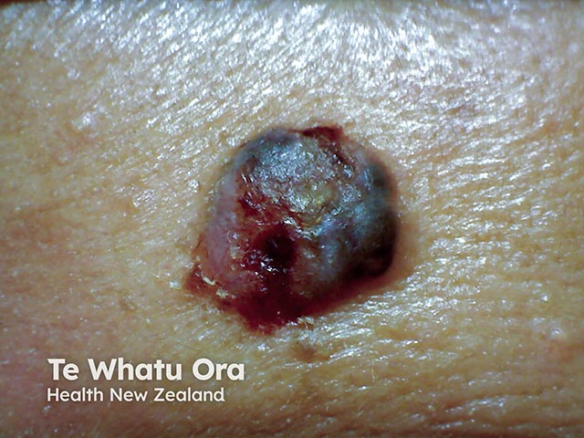
Nodular ulcerated basal cell carcinoma in HIV infection
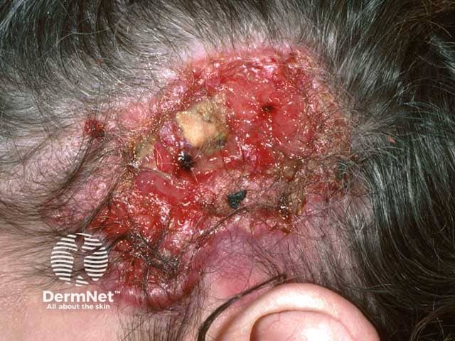
Giant neglected basal cell carcinoma ulcerated down to the skull
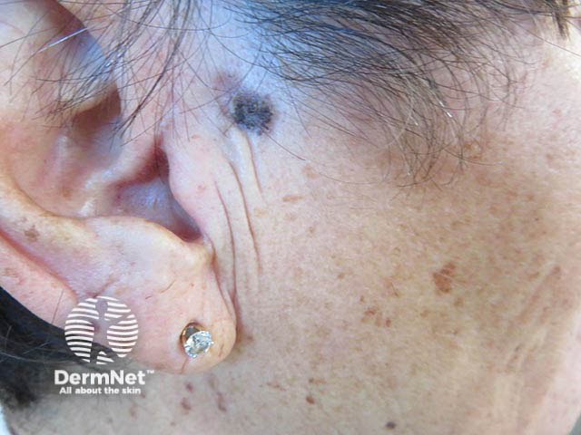
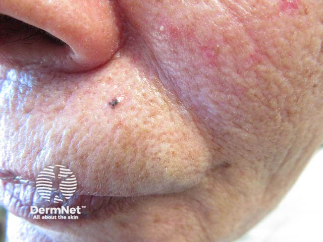
A small pigmented basal cell carcinoma on the upper lip (BCC-patient1)
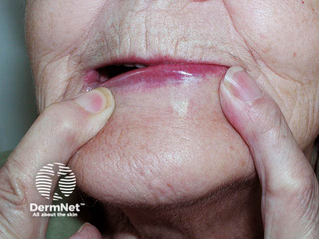
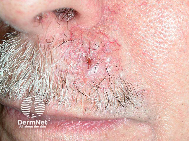
A morphoeic basal cell carcinoma on the left lower lip - it is more easily seen when the skin is stretched
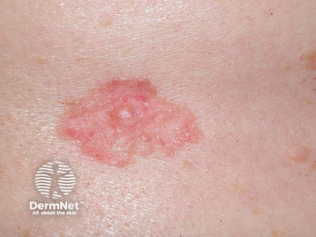
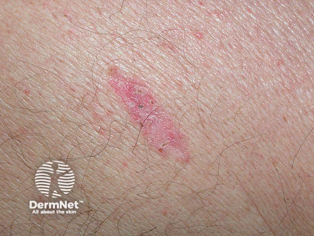
A superficial basal cell carcinoma - they are often pink and have an irregular thread-like edge
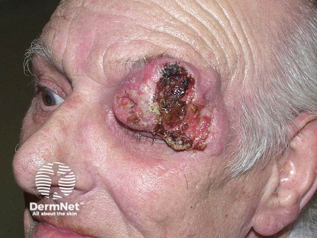
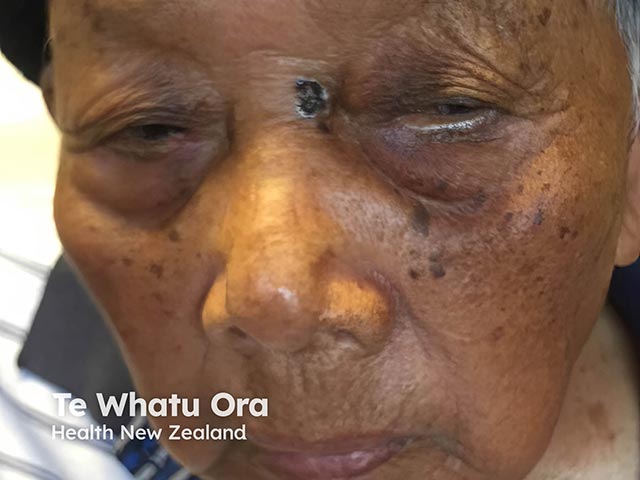
An advanced and neglected basal cell carcinoma on the eyelid
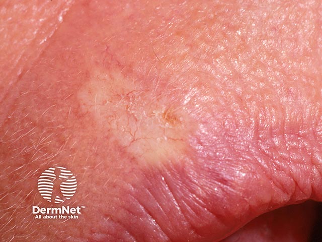
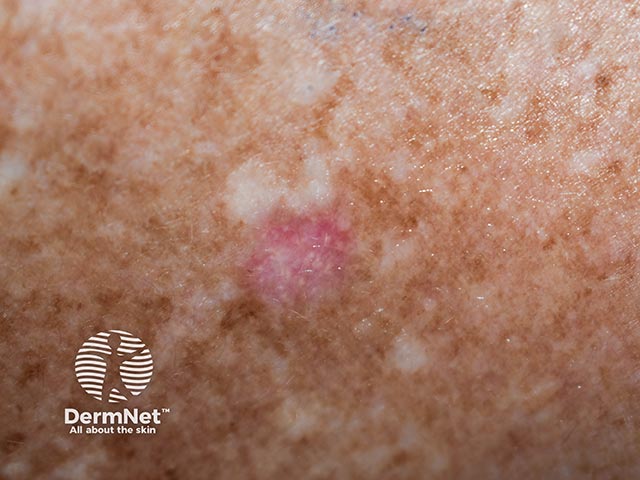
A slowly-spreading pink and well-marginated superficial basal cell carcinoma
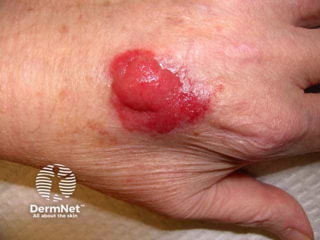
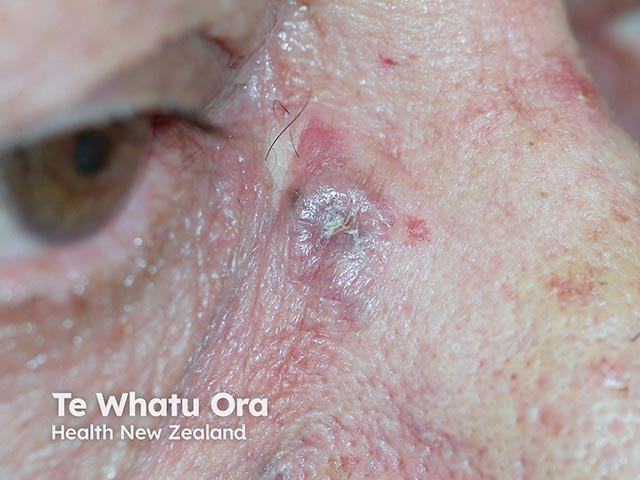
A nodulocystic basal cell carcinoma on the upper lip - the enlarged vessels coursing over the pearly tumour are characteristic
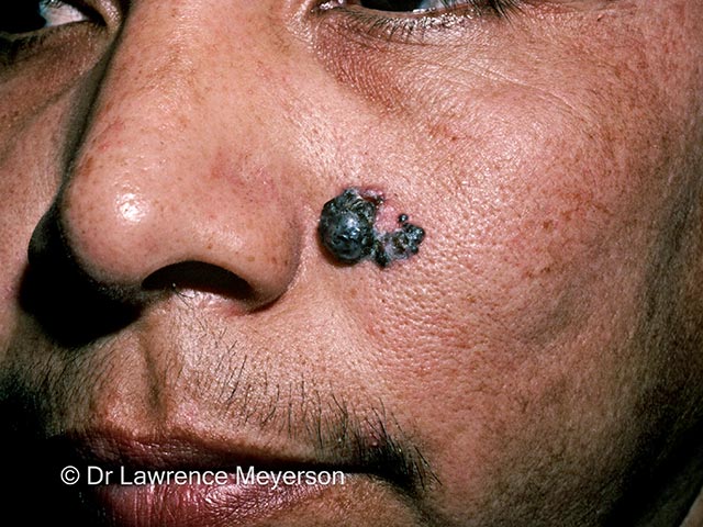
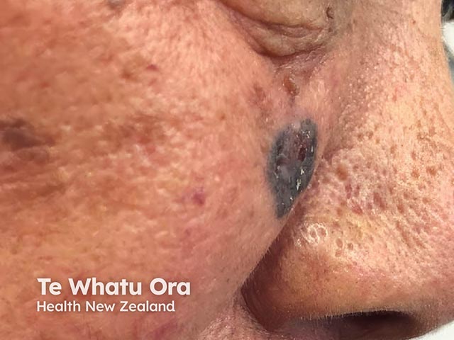
A nodular basal cell carcinoma on the upper nasolabial fold (BCC-patient2)
A nodular basal cell carcinoma on the upper nasolabial fold (BCC-patient2)
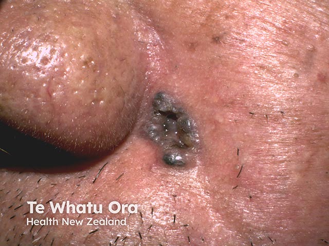
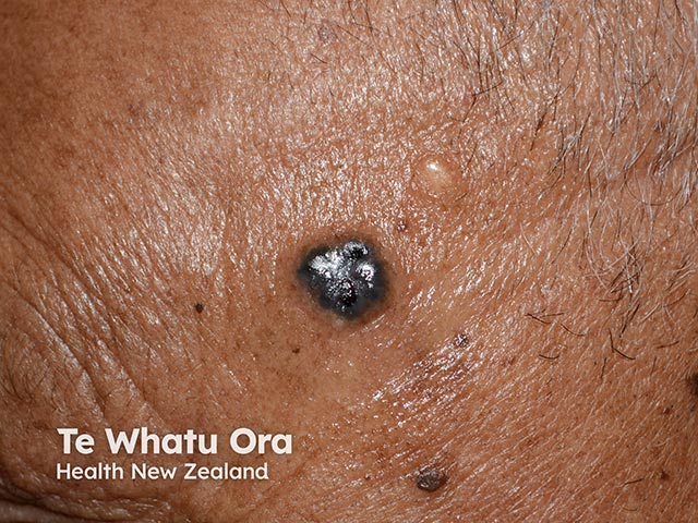
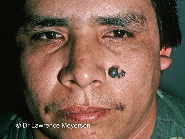
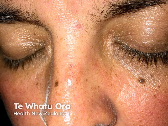
A small pigmented basal cell carcinoma in skin of colour (BCC-patient3)
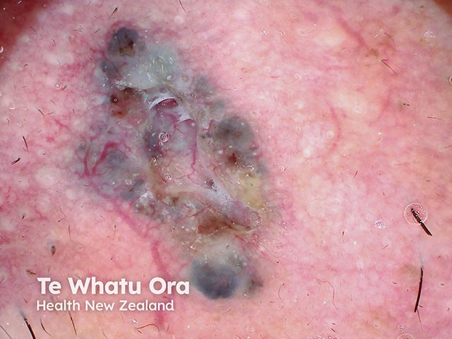
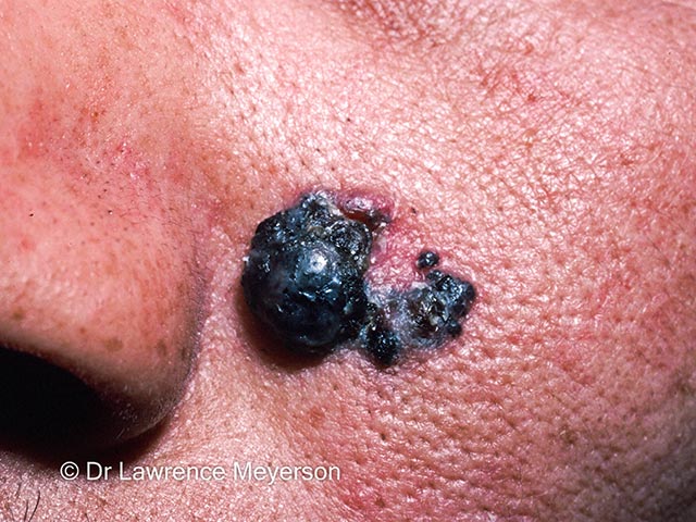
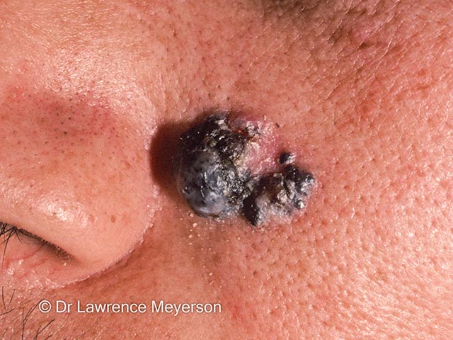
An ulcerated pigmented basal call carcinoma at scalp margin (BCC-patient3)
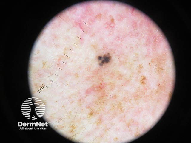
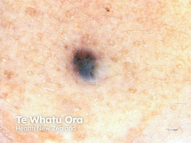
A pigmented basal cell carcinoma in skin of colour on dermoscopy - note the telangiectasia and blue-grey blobs (BCC-patient4)
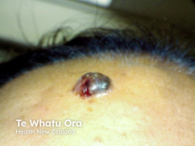
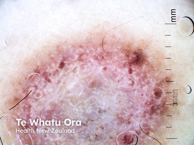
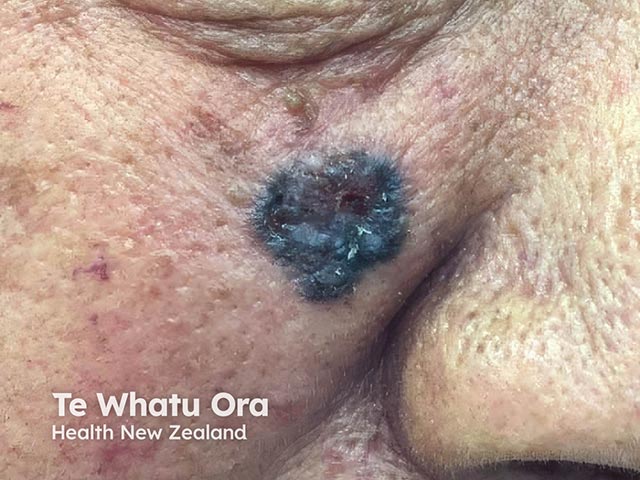
A nodular pigmented basal cell carcinoma in a Māori patient (BCC-patient5)
A nodular pigmented basal cell carcinoma in a Māori patient (BCC-patient5)
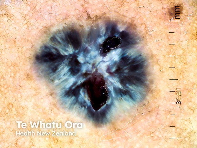
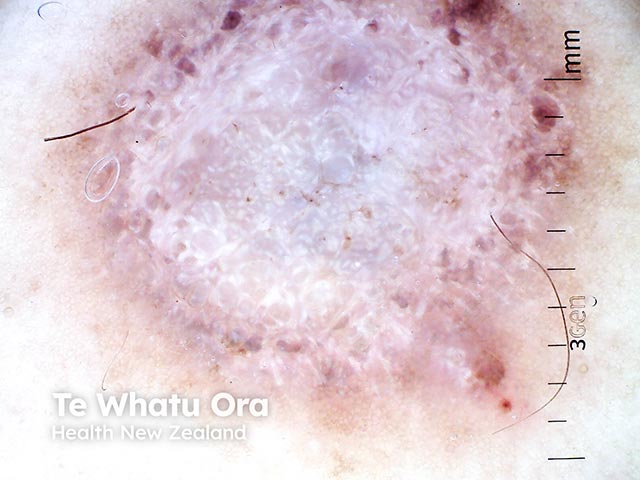
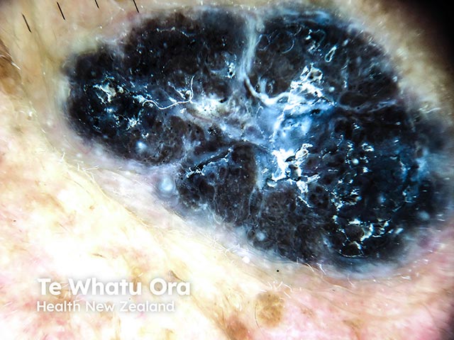
Dermoscopic image of a pigmented basal cell carcinoma in skin of colour - note the spoke-wheel pattern (BCC-patient5)
Superficial basal cell carcinoma arising in a dermatofibroma on the calf (BCC-patient6)
A superficial basal cell carcinoma arising over a dermatofibroma (BCC-patient6)
A superficial basal cell carcinoma arising over a dermatofibroma (BCC-patient6)
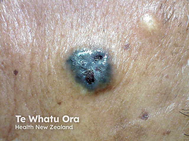
A pigmented nodular basal cell carcinoma in skin of colour (BCC-patient7)
A pigmented nodular basal cell carcinoma in skin of colour (BCC-patient7)
A pigmented nodular basal cell carcinoma in skin of colour (BCC-patient7)
A pigmented nodular basal cell carcinoma in skin of colour (BCC-patient7)
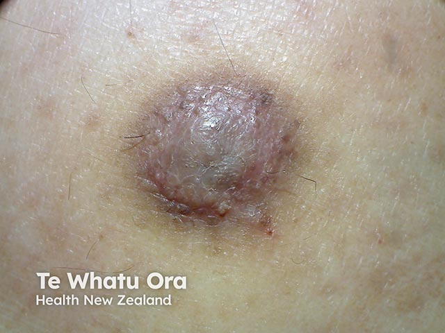
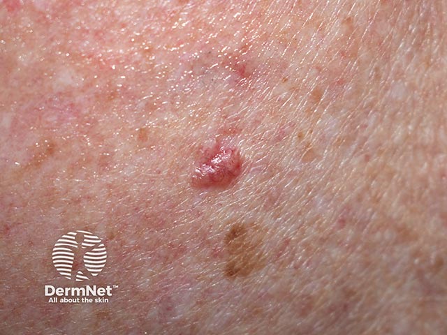
A small nodular basal cell carcinoma in a woman of Indian heritage (BCC-patient8)
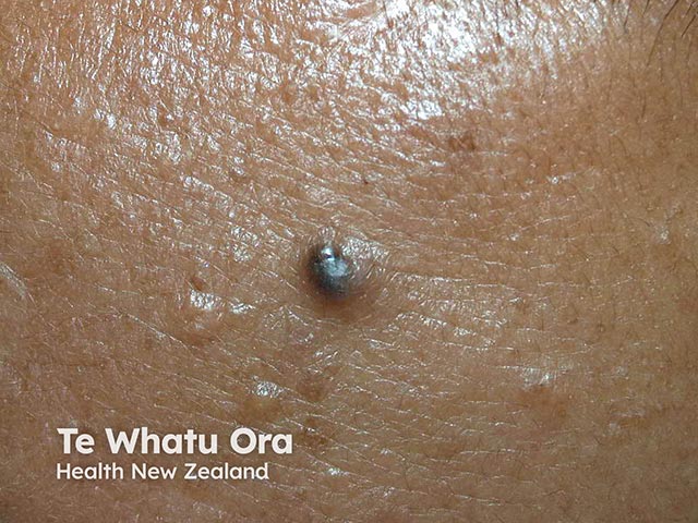
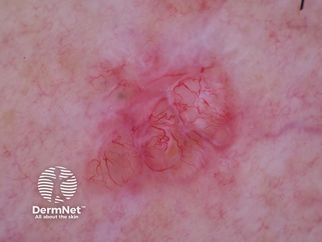
Telangiectatic vessels in a small basal cell carcinoma in skin of colour (BCC-patient8)
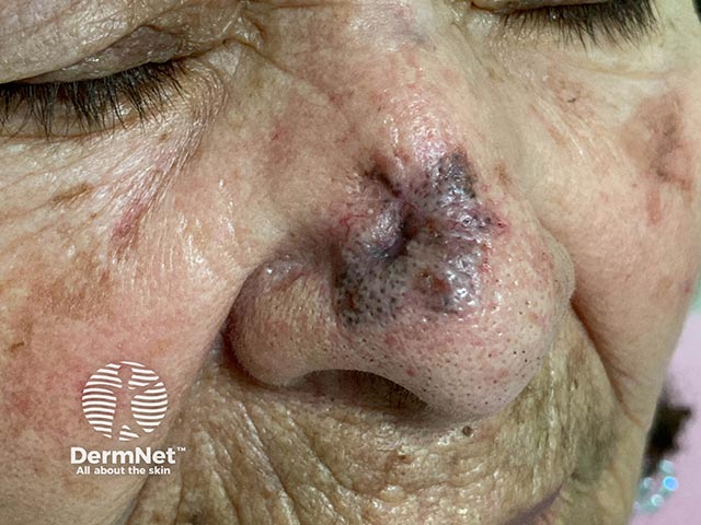
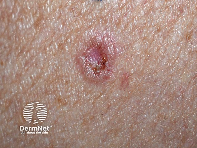
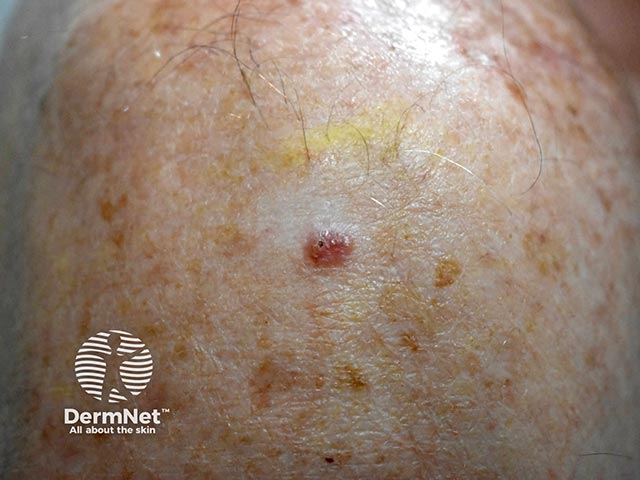
A small basal cell carcinoma on the nose - note the angulated telangiectatic vessels
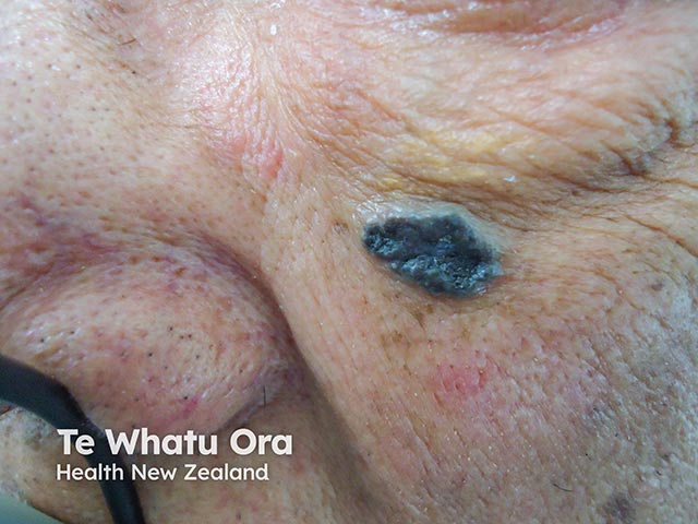
Dermoscopy of a deeply pigmented basal cell carcinoma in a Pacific Islander (BCC-patient9)
A pigmented basal cell carcinoma in an elderly South East Asian woman (BCC-patient11)
A morphoeic basal cell carcinoma on the lip - note the branched telangiectasia within the scar-like lesion
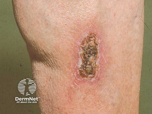
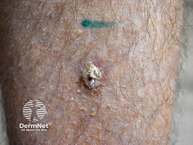
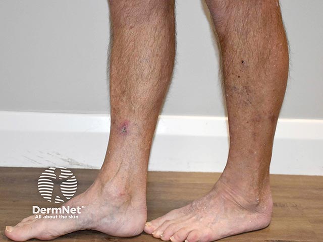
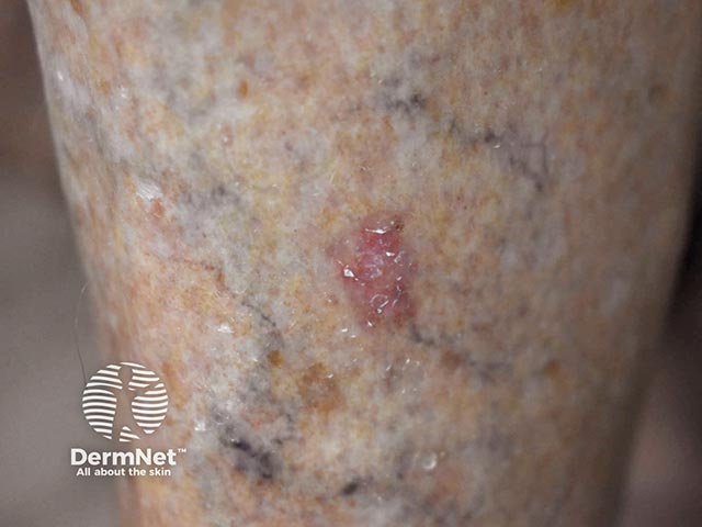
A medial calf basal cell carcinoma - it may mimic a venous ulcer
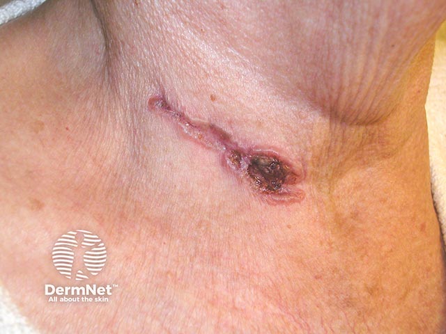
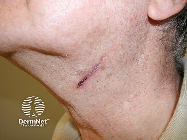
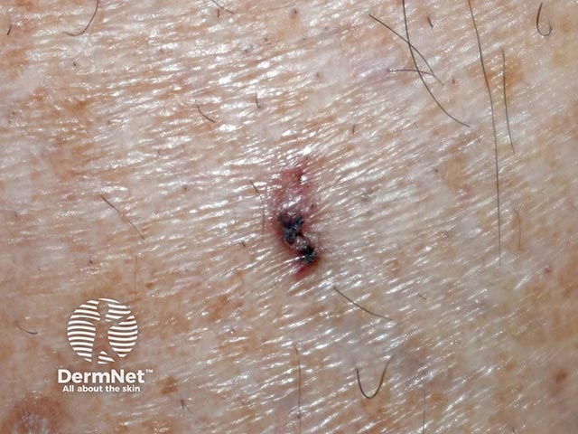
A linear basal cell carcinoma on the neck
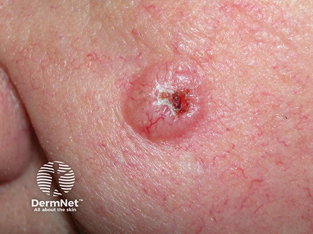
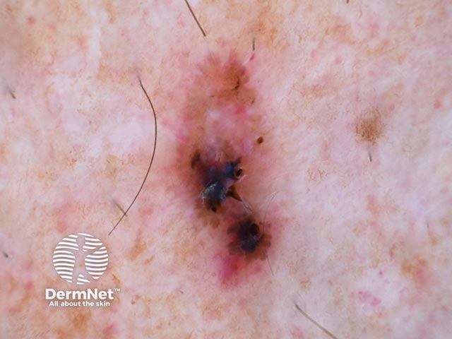
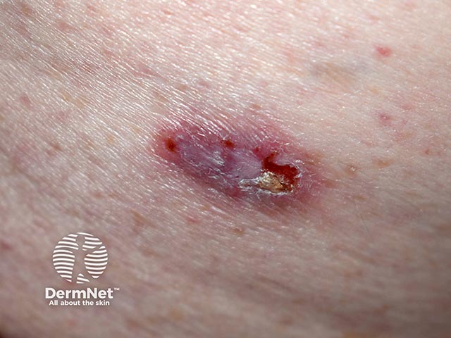
A typical ulcerated nodular basal cell carcinoma - note the pearly appearence and broken telangiectatic vessels
A linear basal cell carcinoma on the neck
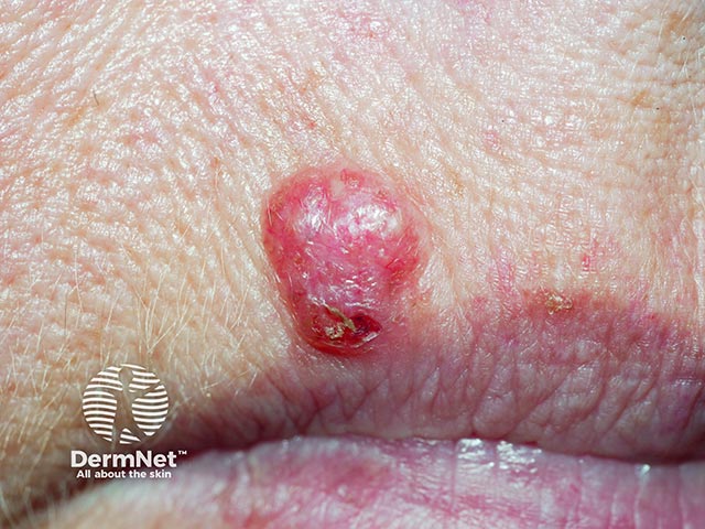
A nodular basal cell carcinoma on the lip - note the inferior ulceration
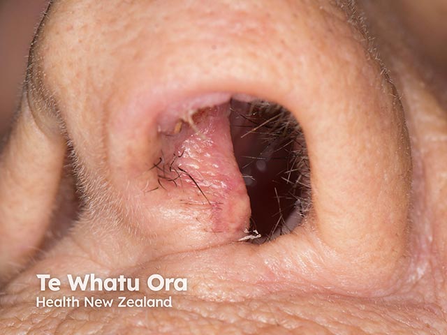
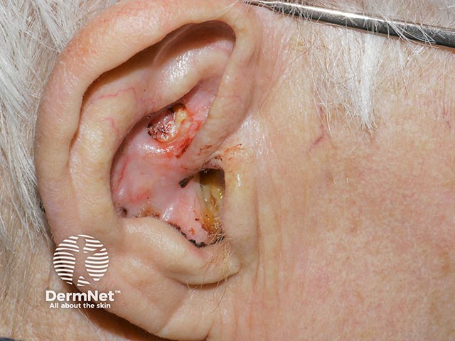
An ulcerated basal cell carcinoma in the sulcus of the pinna
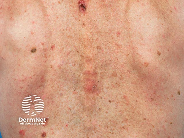
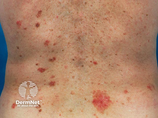
Multiple superficial basal cell carcinomas after spinal radiotherapy (BCC-patient13)
Multiple superficial basal cell carcinomas after spinal radiotherapy (BCC-patient13)
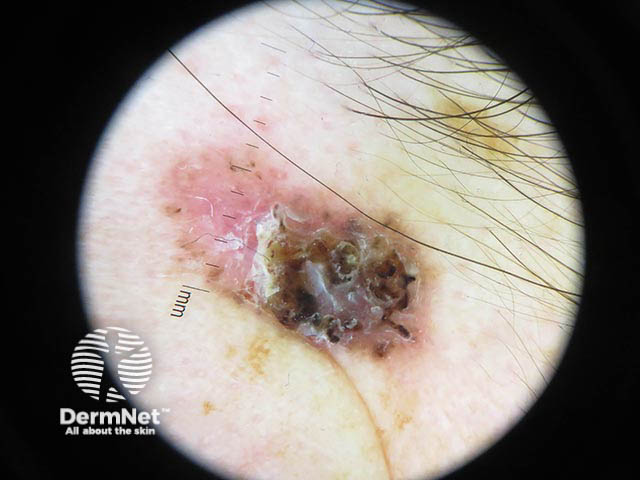
A pigmented basal cell carcinoma on dermoscopy - note the broken telangiectasia and pigment flecks (BCC-patient14)
A pigmented basal cell carcinoma on dermoscopy - note the broken telangiectasia and pigment flecks (BCC-patient14)
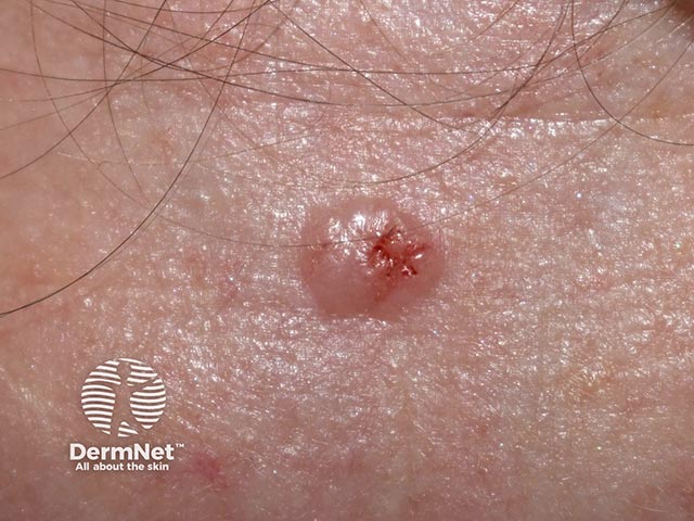
A typical basal cell carcinoma - note the pearly raised edge, crusting, and depressed centre (BCC-patient15)
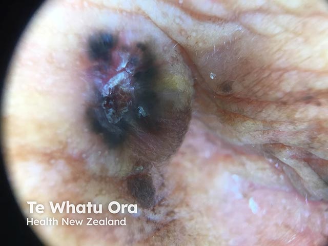
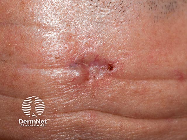
Close-up of a basal cell carcinoma - crust is obliterating some of the diagnostic features (BCC-patient16)
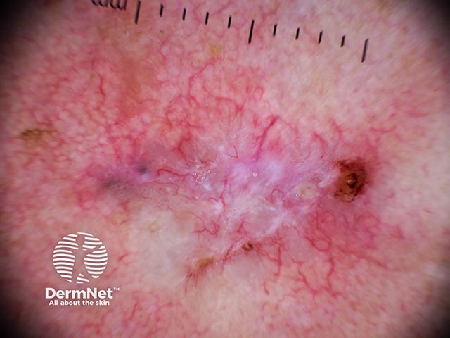
The glassy periphery and central ulceration revealed after removing the crust (BCC-patient16)
Dermoscopy shows the broken arborising telangiectasia and central featureless sclerosis (BCC-patient16)
Dermoscopy shows the broken arborising telangiectasia and central featureless sclerosis (BCC-patient16)
Basal cell carcinoma close-up with scale and crust obliterating the diagnostic features (BCC-patient16)
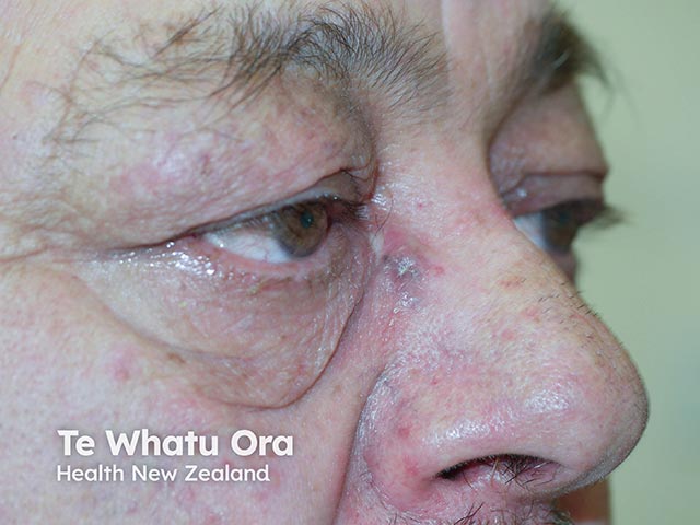
A basal cell carcinoma showing central atrophy and a pigmented periphery (BCC-patient18)
Dermoscopy of a basal cell carcinoma - pigmented clods, fine telangiectatic vessels, and central white-cream atrophy (BCC-patient18)
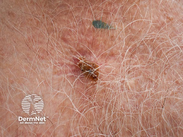
A nodular basal cell carcinoma on the forehead - note the raised pearly edge, telangiectasia, and central depression (BCC-patient19)
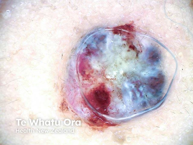
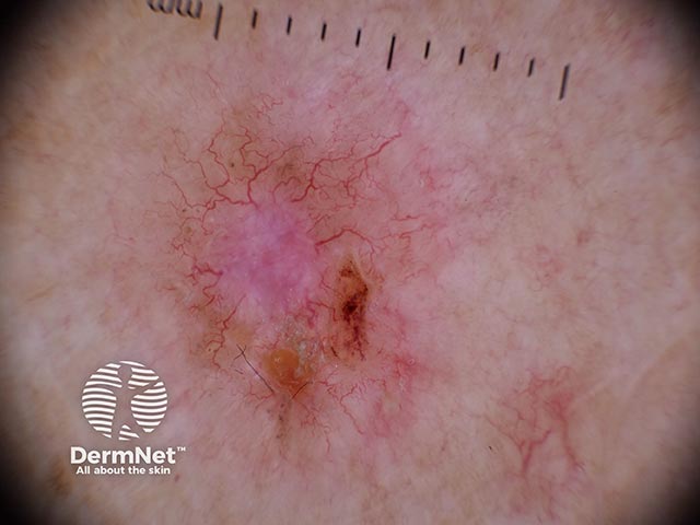
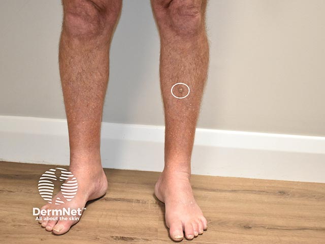
Dermoscopic image of a basal cell carcinoma on the lower leg (BCC-patient19)
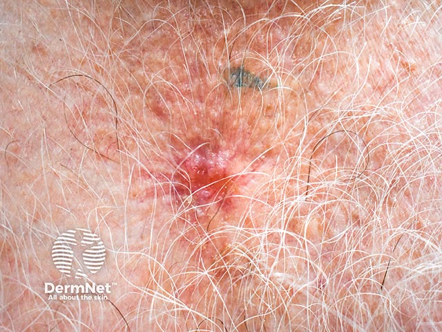
Dermoscopy shows white-pink areas and telangiectatic vessels (BCC-patient19)
Dried blood obscures many diagnostic dermatoscopic features (BCC-patient19)
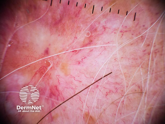
Dermoscopy shows white structureless areas and fine linear vessels (BCC-patient19)
Dermoscopy showing crust and scale with pink-white area above (BCC-patient19)
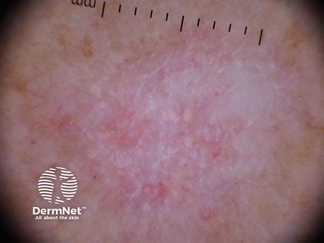
Dermoscopy shows white-grey areas and linear and branched telangiectasia (BCC-patient20)
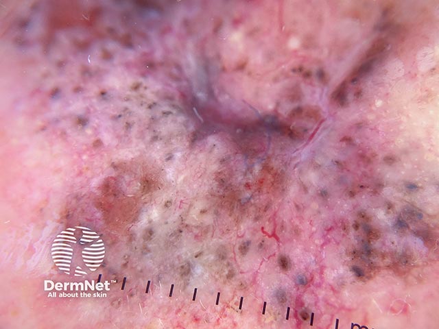
Dermoscopy showing clods of pigment and linear radial vessels (BCC-patient21)
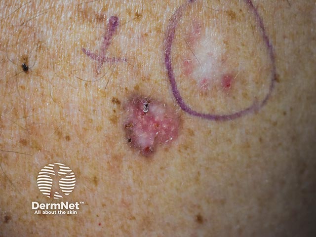
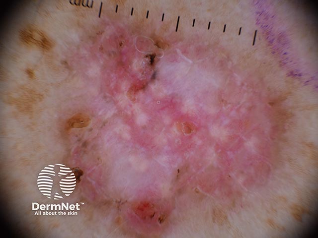
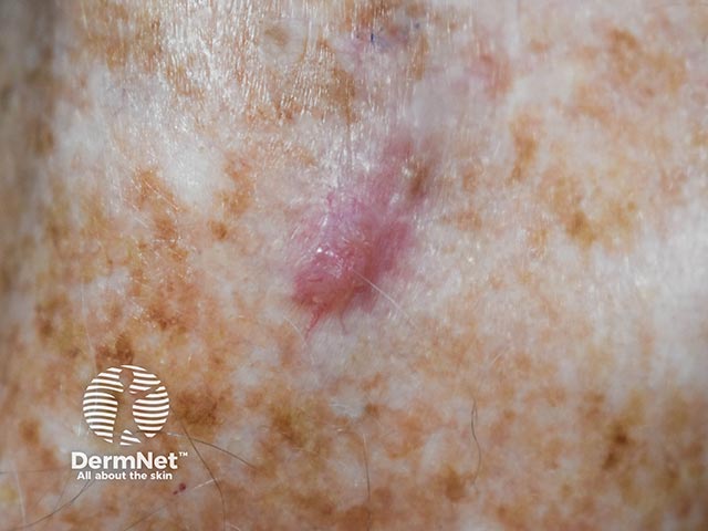
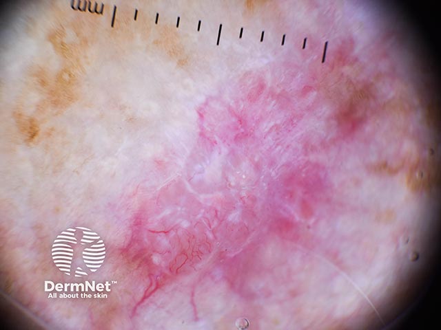
Dermoscopy of a basal cell carcinoma on the leg - note pigmentation and pink areas (BCC-patient22)
Dermoscopy of a basal cell carcinoma on the leg - note fine vessels and clumps of pigment at the upper pole (BCC-patient23)
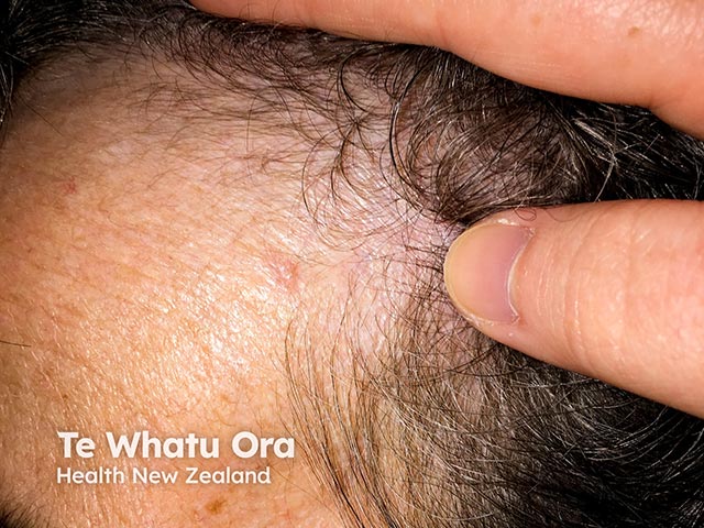
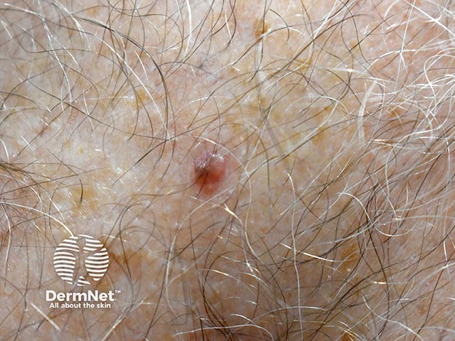
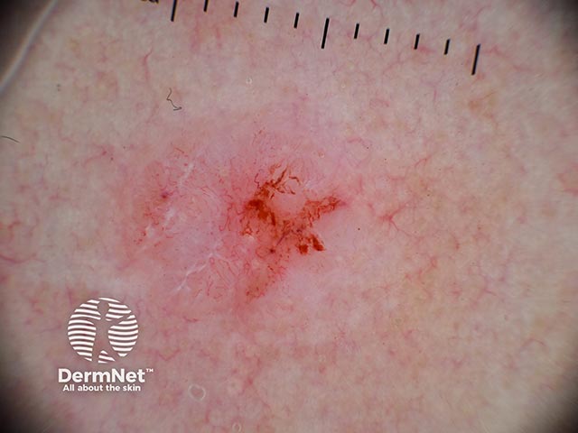
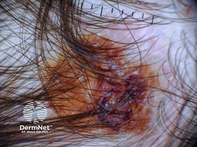
A basal cell carcinoma on the scalp - diagnostic features are obscured by the overlying crust (BCC-patient25)
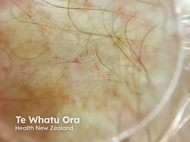
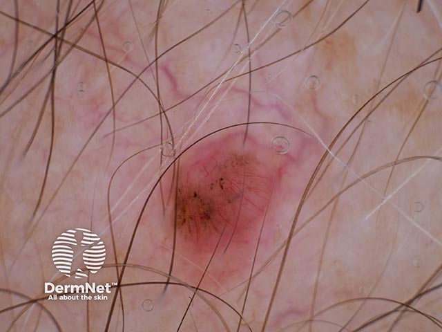
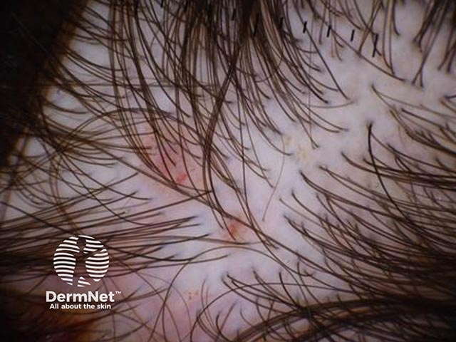
Dermoscopic image of superior satellite plaques from large infiltrating basal cell carcinoma (BCC-patient25)
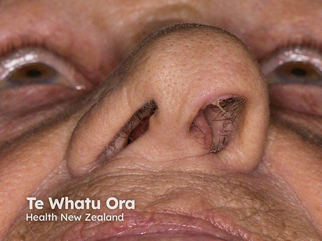
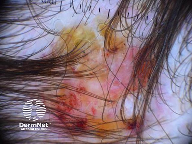
Appearances of a basal cell carcinoma on the scalp after removal of the crust (BCC-patient25)
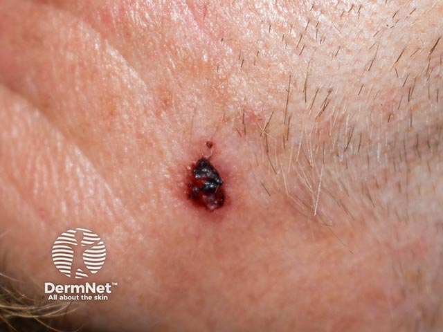
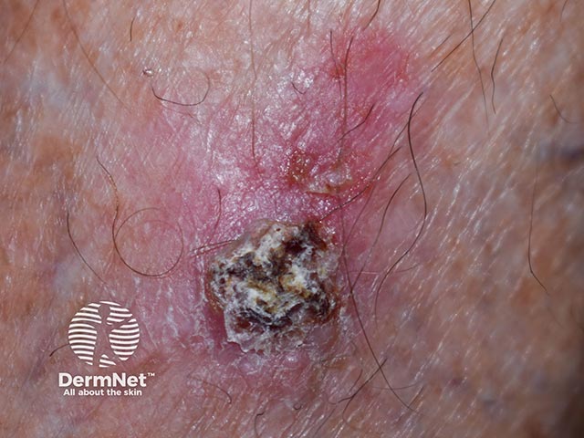
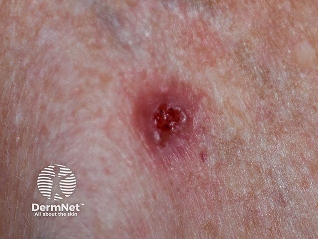
An ulcerated basal cell carcinoma - note the pearly raised edge (BCC-patient26)
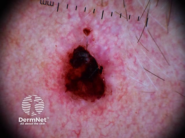
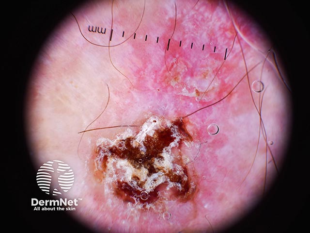
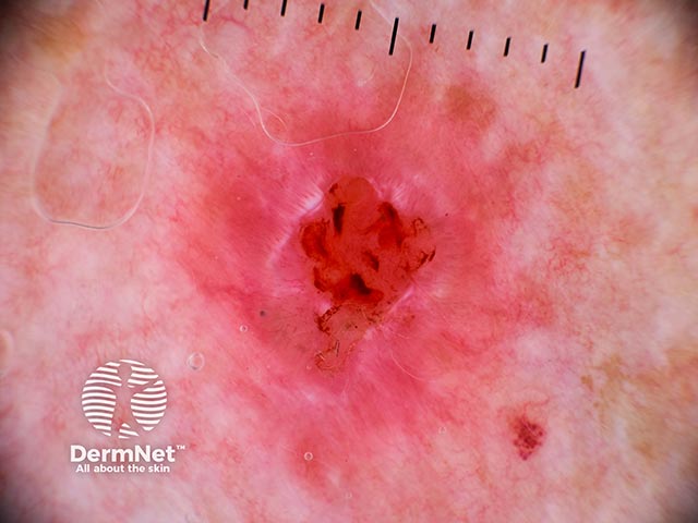
Basal cell carcinoma - note adherent fibres in the adherent serous exudate (BCC-patient26)
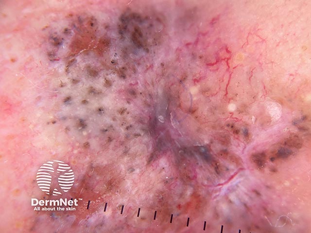
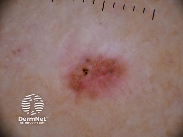
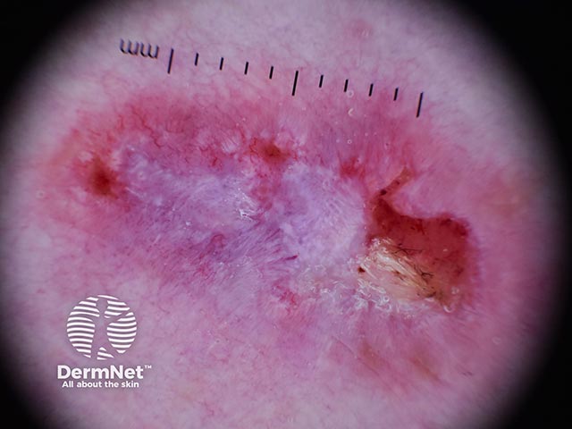
Dermoscopy of a basal cell carcinoma on the leg - note ulceration and peripheral telangiectasia and grey-white central islands (BCC-patient27)
Go to basal cell carcinoma topic page
Basal cell carcinoma histopathology images
Go to basal cell carcinoma pathology page
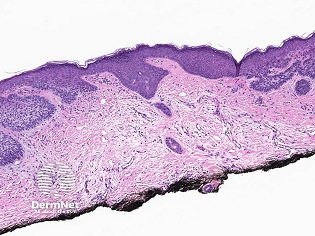
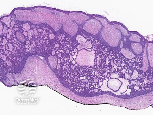
A superficial basal cell carcinoma - buds of atypical basaloid cells bud down towards the dermis
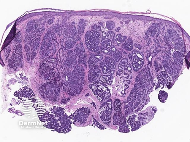
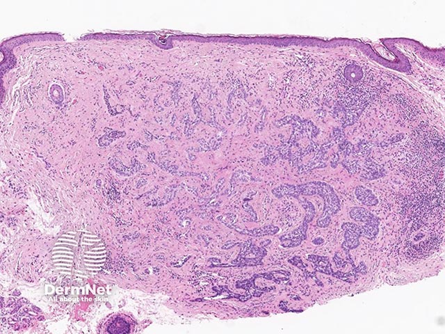
A nodular basal cell carcinoma - nodules of dark basaloid cells can be seen with a surrounding stroma
A nodular basal cell carcinoma - the peripheral palisading of the basaloid cells can be appreciated
A pigmented basal cell carcinoma - flecks of melanin pigment can be seen within the stroma of the tumour
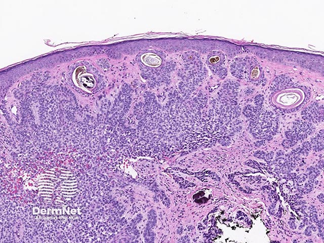
An infiltrative basal cell carcinoma - strands and buds of basaloid cells can be seen deep into the dermis
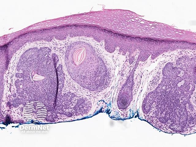
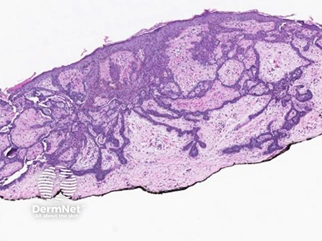
A fibroepithelioma of Pinkus - net-like interconnecting strands of basaloid cells and a vascular stroma are noted
A fibroepithelioma of Pinkus - interconnecting strands of basaloid cells interspersed with vascularised stroma
Go to basal cell carcinoma pathology page