Main menu
Common skin conditions

NEWS
Join DermNet PRO
Read more
Quick links
Go to the calcinosis cutis topic page
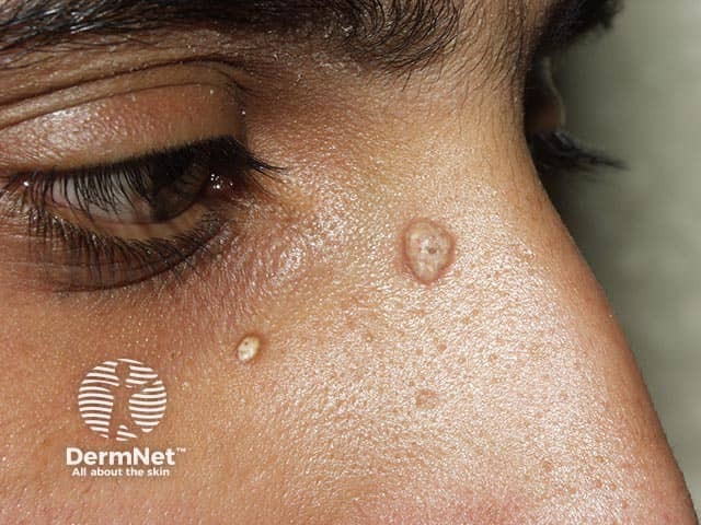
A subepidermal calcified nodule on the nose
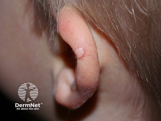
A subepidermal calcified nodule on the ear - note the typical chalky appearence
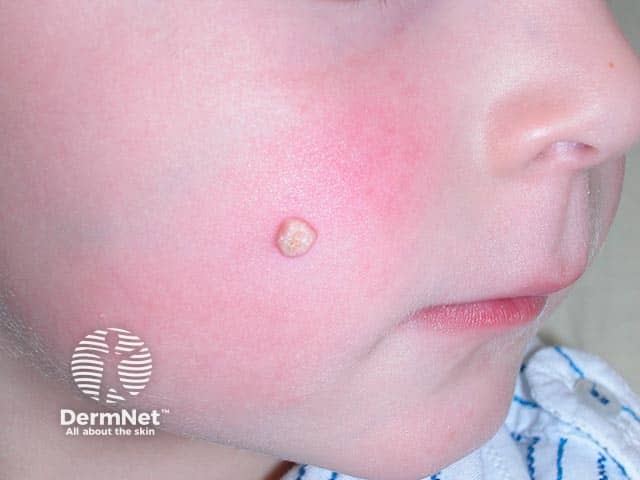
A subepidermal calcified nodule on the cheek - cheeks and ears are the most frequent location
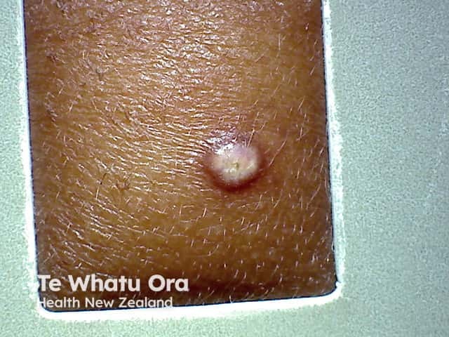
A subepidermal calcified nodule on the cheek (CC-patient1)
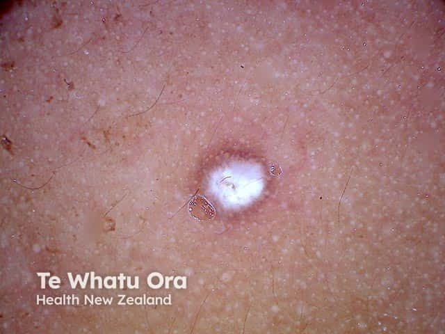
A non-polarised dermatoscopic image of a subepidermal calcified nodule on the face of a child (CC-patient1)

A polarised dermatoscopic image of a subepidermal calcified nodule on the face of a child (CC-patient1)
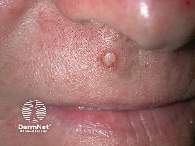
A subepidermal calcified nodule on the lip of an adult - they are more common in children
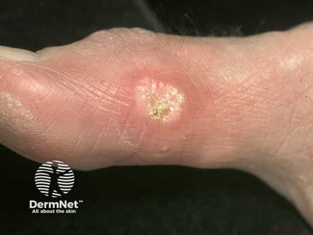
Idiopathic cutaneous calcification on the side of the foot
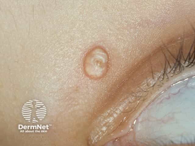
A subepidermal calcified nodule on the lid
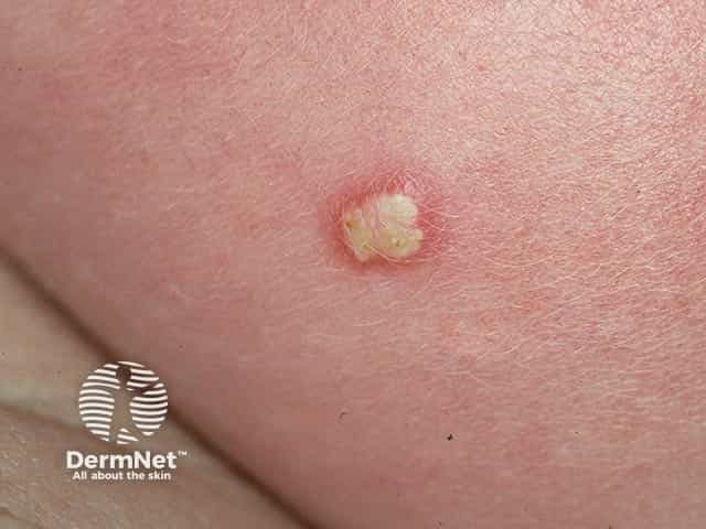
A subepidermal calcified nodule on the cheek
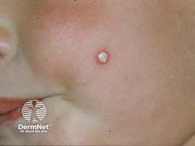
The typical chalky white appearence of a supepidermal calcified nodule on the cheek
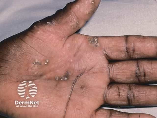
Multiple lesions of calcinosis cutis on the palm (CC-patient2)
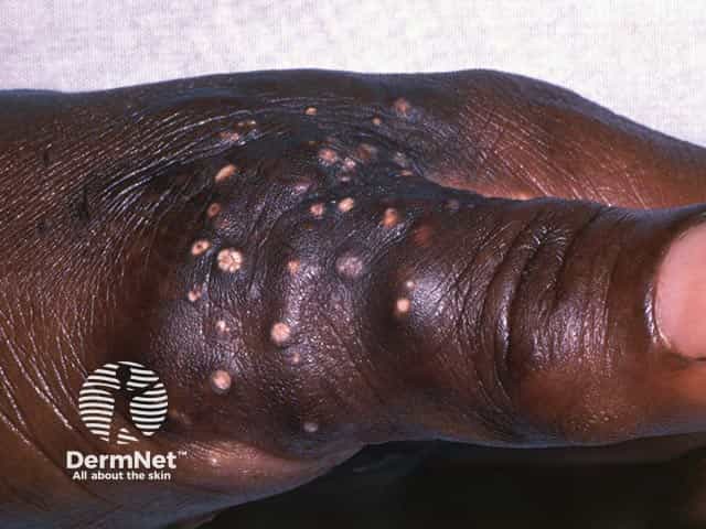
Multiple lesions of calcinosis cutis on the base of the thumb (CC-patient2)
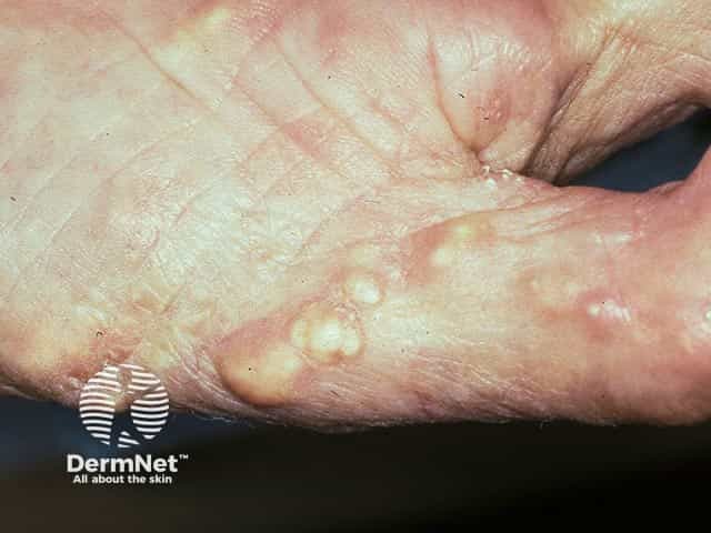
Calcinosis cutis on the thumb - most commonly seen in CREST syndrome (CC-patient3)
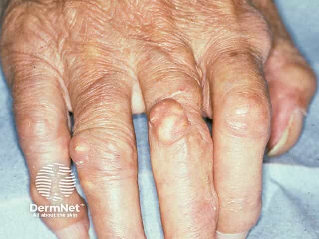
Calcinosis cutis on the fingers - most commonly seen in CREST syndrome (CC-patient3)
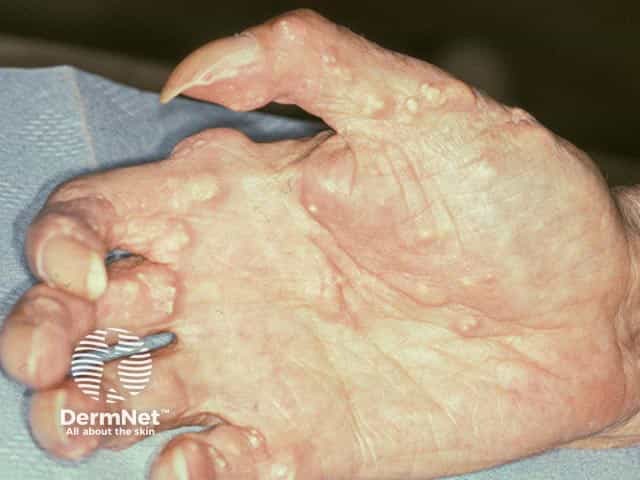
Calcinosis cutis on the fingers - most commonly seen in CREST syndrome (CC-patient3)
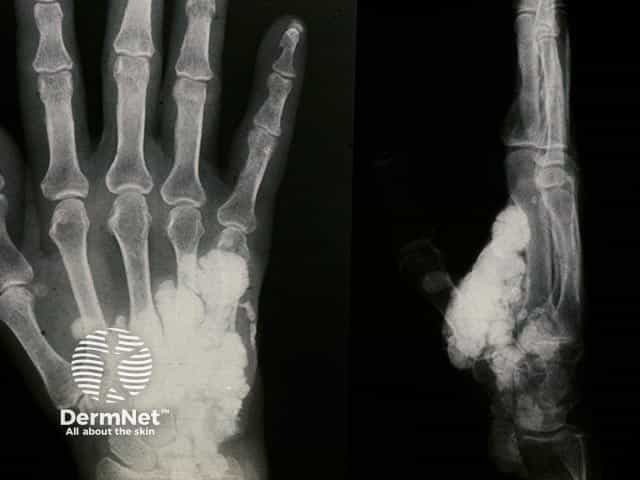
Radiograph of the hand in extensive calcinosis cutis showing palmar calcification
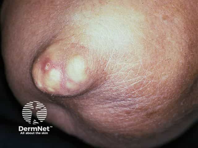
Nodular calcinosis cutis on the elbow
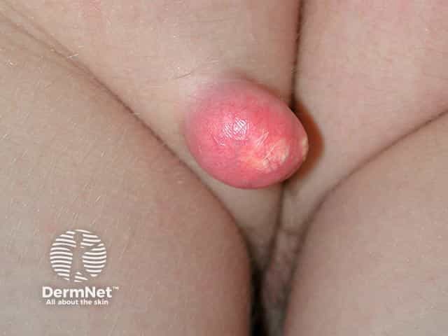
A unusually large subepidermal calcified nodule in an unusual location on the buttock
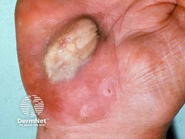
A large area of calcinosis cutis on the heel of the palm
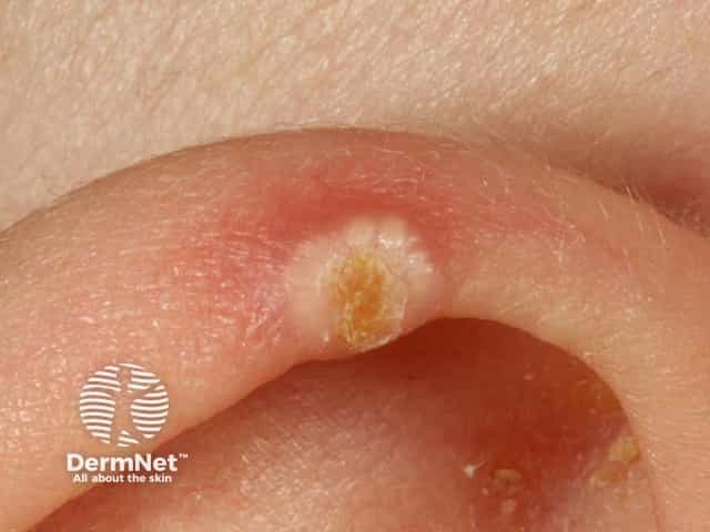
A chalky-white gritty nodule on the ear due to a subepidermal calcified nodule
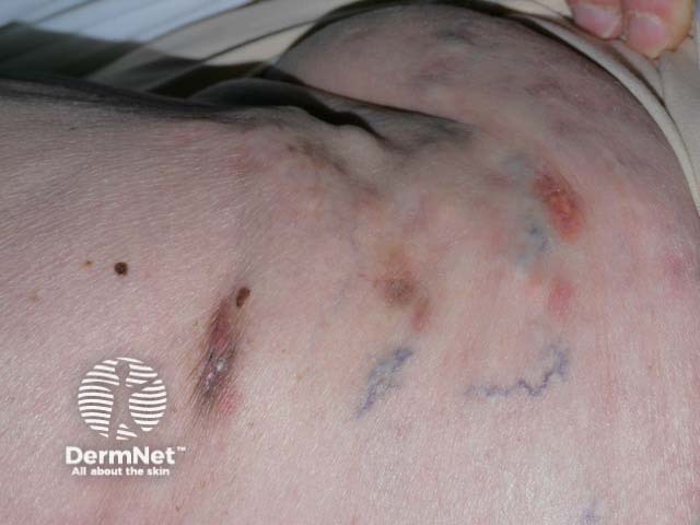
Extensive calcinosis cutis over the chest wall due to underlying dermatomyositis. Overlying ulceration and calcium extrusion has occurred in places