The deposition of calcium in the skin, subcutaneous tissue, muscles and visceral organs is known as calcinosis. This condition commonly occurs in the skin, where it is known as calcinosis cutis or cutaneous calcification.
Calcinosis cutis is classified into four major types.
Dystrophic calcinosis cutis occurs in an area where there is damaged, inflamed, neoplastic or necrotic skin. Tissue damage may be from mechanical, chemical, infectious or other factors. Normal serum calcium and phosphate levels exist. Conditions that can cause dystrophic calcinosis cutis may include:
Metastatic calcinosis cutis occurs in the setting of abnormal calcium and phosphate metabolism and is often associated with hypercalcaemia and/or hyperphosphataemia. Conditions that can cause metastatic calcinosis cutis may include:
Idiopathic calcinosis cutis generally occurs in the absence of any known tissue injury or systemic metabolic defect. Calcification is usually localised to one general area.
Iatrogenic calcinosis cutis arises secondary to a treatment or procedure, for example, parenteral administration of calcium or phosphate, and calcium deposition in newborns from repeated heel sticks.
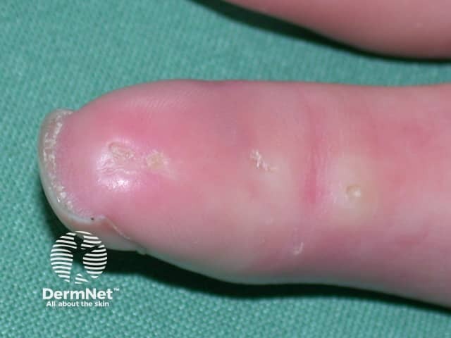
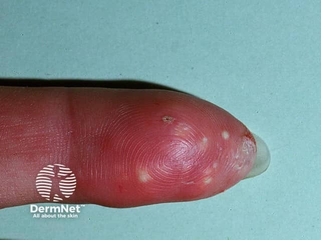
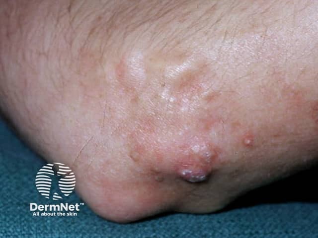
The signs and symptoms of calcinosis cutis vary according to the underlying cause. In many cases, the lesions gradually develop and are often symptomless. The lesions usually appear as firm, whitish/yellowish papules, plaques or nodules on the surface of the skin. A solitary lesion may develop, although multiple lesions are more common. Lesions may become tender and ulcerate, discharging chalk-like creamy material consisting mainly of calcium phosphate with a small amount of calcium carbonate.
Fingertip lesions may be painful, while lesions at other sites may restrict joint mobility and limit movement due to stiffening of the skin. In severe cases, cutaneous gangrene may occur.
Laboratory tests are performed to determine any metabolic abnormalities that may give rise to elevated calcium and phosphate levels. Radiological examinations including plain film x-ray, CT scanning and bone scintigraphy are useful in demonstrating the extent of tissue calcification.
Biopsy of cutaneous lesions is used to confirm the diagnosis. On histology of calcinosis cutis, granules and deposits of calcium are seen in the dermis, often with a surrounding foreign-body giant cell reaction. Calcium deposits may also be found in subcutaneous tissue.
The underlying cause of calcinosis cutis should be identified and treated accordingly. Medical therapy may be used to help relieve symptoms of the condition but are generally of limited and variable benefit.
Medications that may be tried include:
Surgical or carbon dioxide removal of lesions is indicated when they:
Because surgical trauma may stimulate further calcification, it may be best to excise a small site before going ahead with a large excision. Recurrence is common after excision.
Depending on the underlying cause, a multidisciplinary team of physicians including nephrologist, rheumatologist, and haematologist may be needed to manage the condition.

