Main menu
Common skin conditions

NEWS
Join DermNet PRO
Read more
Quick links
Go to the nodular melanoma topic page
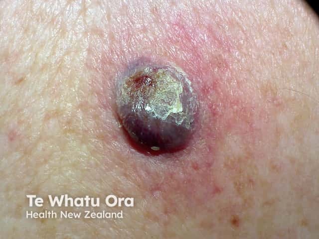
An ulcerated and crusted purple-red rapidly growing lesion on the back - histology showed a nodular melanoma (NM-patient1)
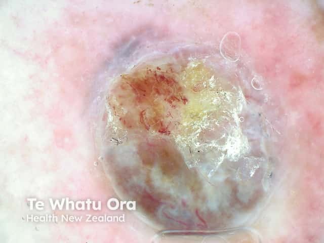
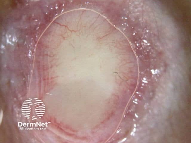
An ulcerated nodular lesion with polymorphous vessels and structureless area - a nodular malignant melanoma (NM-patient1)

A changing unusual mole on the left shoulder - the 'ugly duckling' sign of melanoma (NM-patient2)
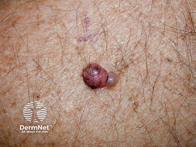
A nodular lesion with a medial satelllite lesion - the larger part is pink with dark brown and black speckles suggesting an amelanotic nodular malignant melanoma (NM-patient2)
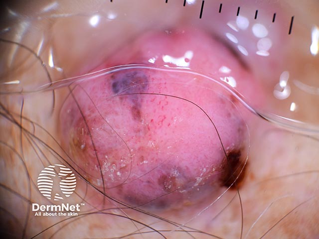
Dermoscopy shows a pink lesion with atypical vessels and speckles of pigment peripherally - it was an 8.3 mm Breslow thickness malignant melanoma (NM-patient2)
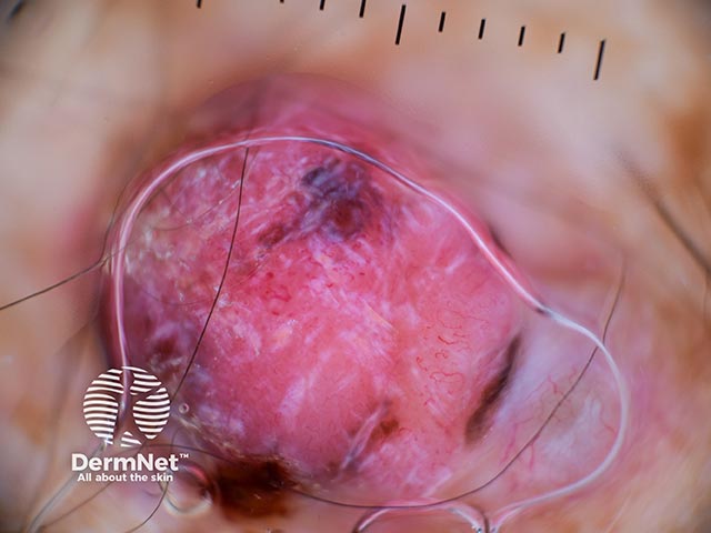
Dermoscopy shows a pink lesion with atypical vessels and speckles of pigment peripherally - it was an 8.3 mm Breslow thickness malignant melanoma (NM-patient2)
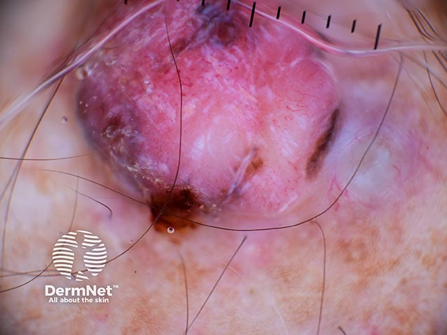
Dermoscopy shows a pink lesion with atypical vessels and speckles of pigment peripherally - it was an 8.3 mm Breslow thickness malignant melanoma (NM-patient2)
Go to the nodular melanoma topic page
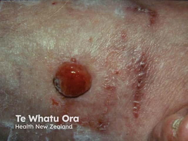
Nodular melanoma
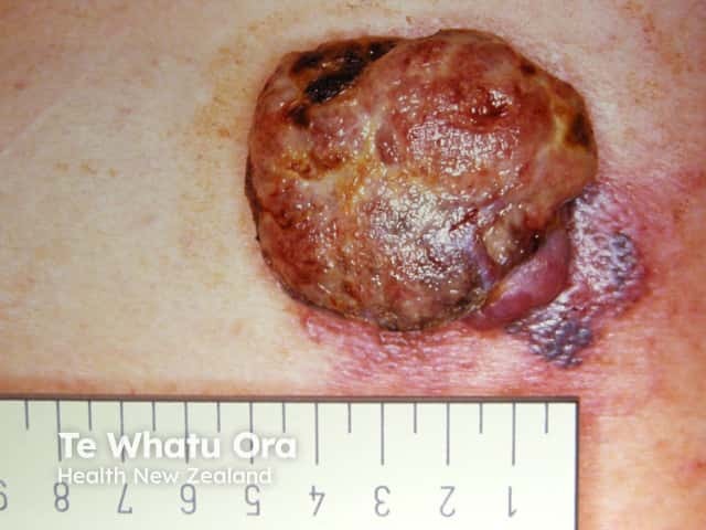
Nodular melanoma

Nodular melanoma
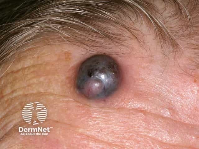
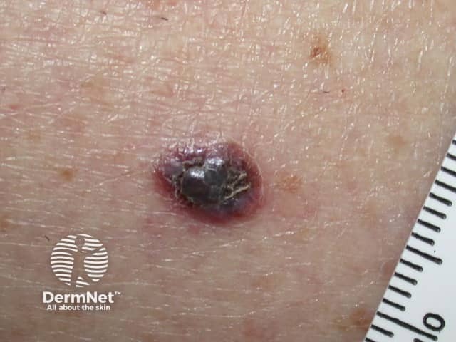
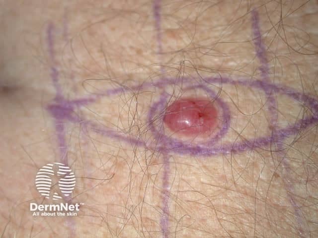
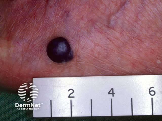
Nodular melanoma
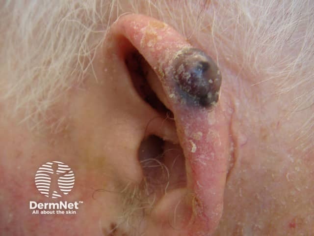
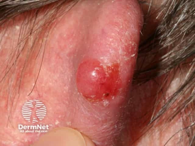
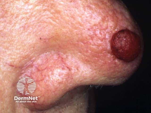
Amelanotic nodular melanoma
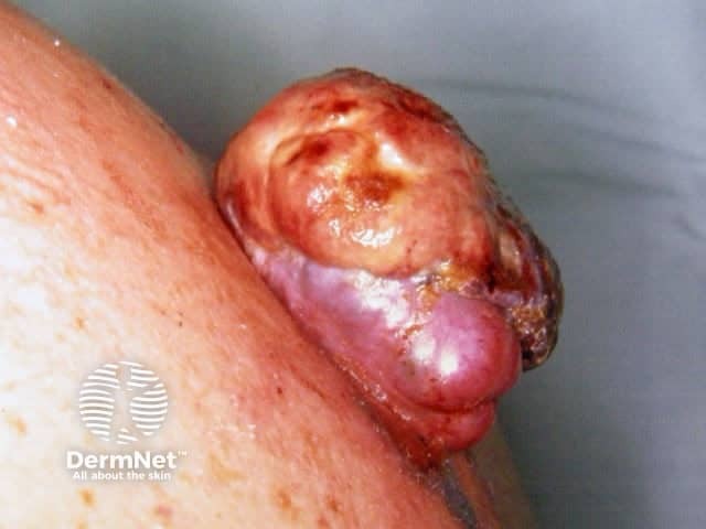
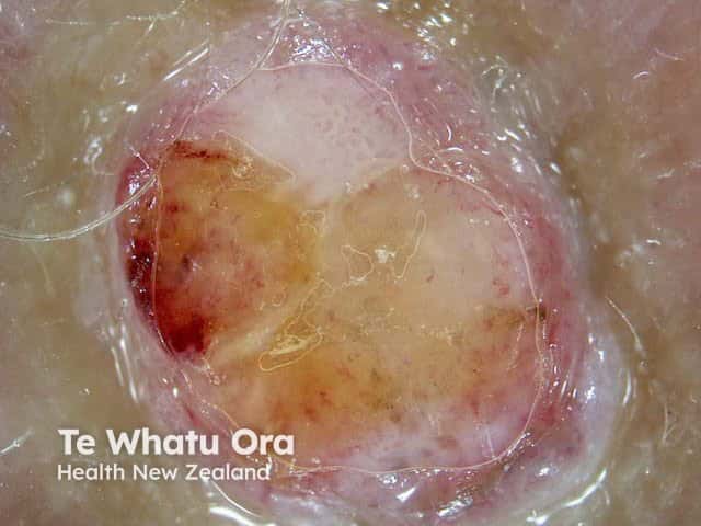
Ulcerated nodular melanoma
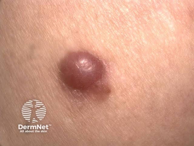
Amelanotic nodular melanoma
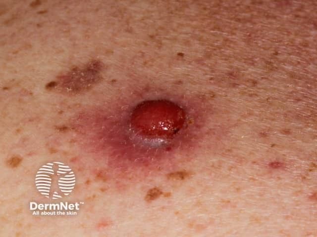
Amelanotic nodular melanoma
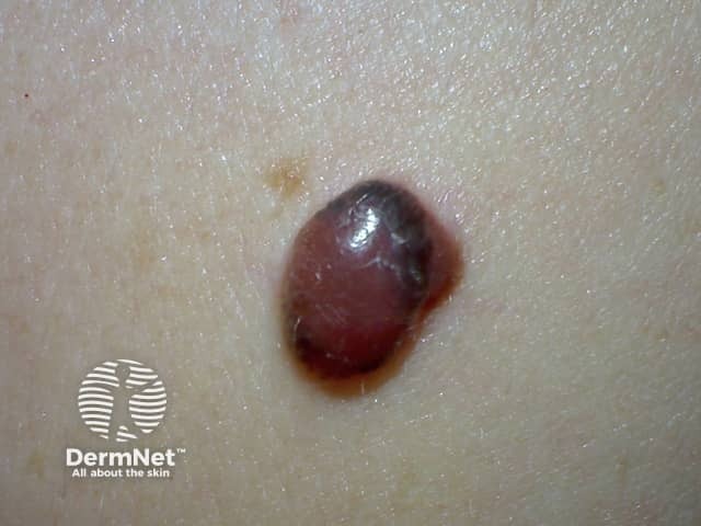
Nodular melanoma

Nodular melanoma
Go to the nodular melanoma topic page
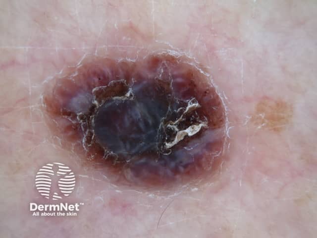
Dermoscopy of nodular melanoma
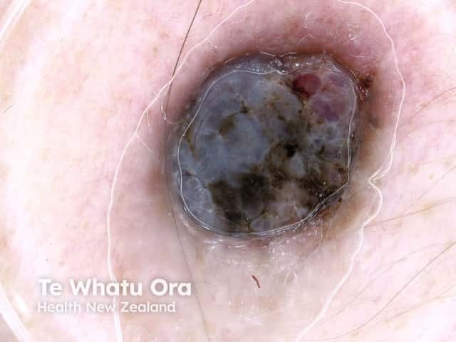
Dermoscopy of nodular melanoma
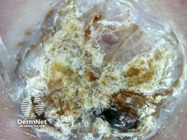
Dermoscopy of a 6 mm thick nodular melanoma
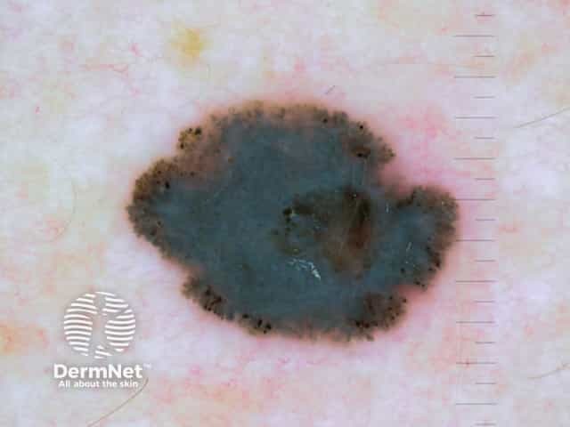
Dermoscopy of nodular melanoma Breslow 0.8 mm
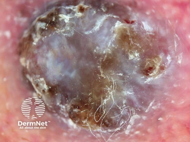
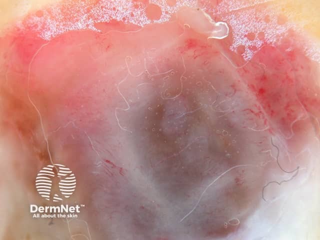
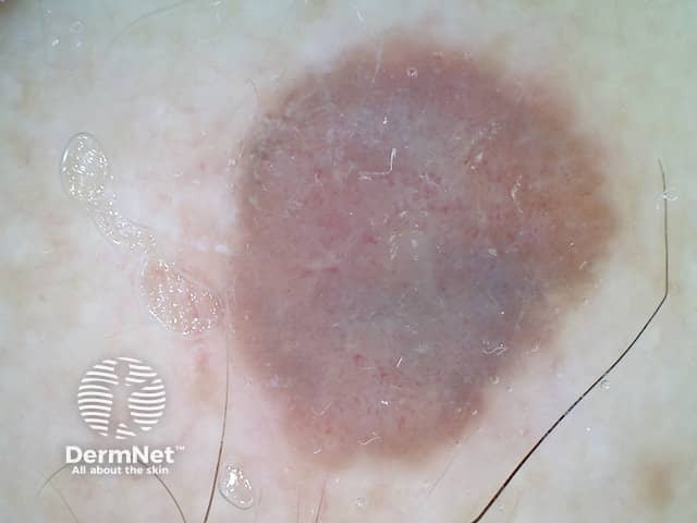
Dermoscopy of nodular melanoma Breslow 4 mm
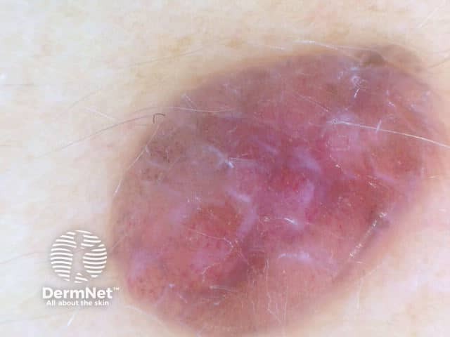
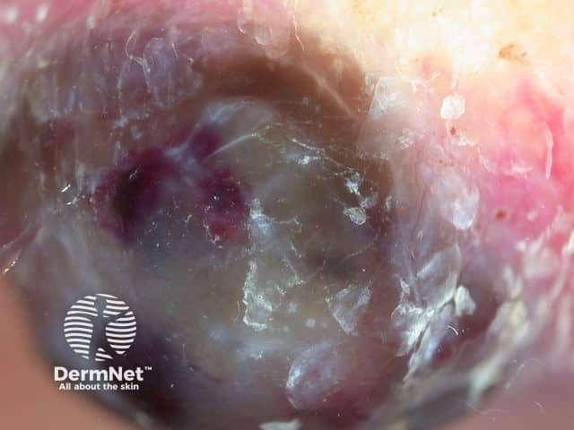
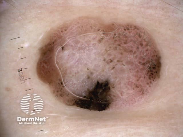
Nodular melanoma, Breslow 2.5 mm, polarised dermoscopy view
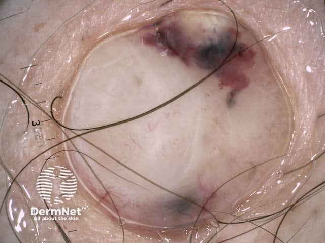
Nodular melanoma, Breslow 7.2 mm, showing changes in the 12 weeks prior to excision, nonpolarised dermoscopy view
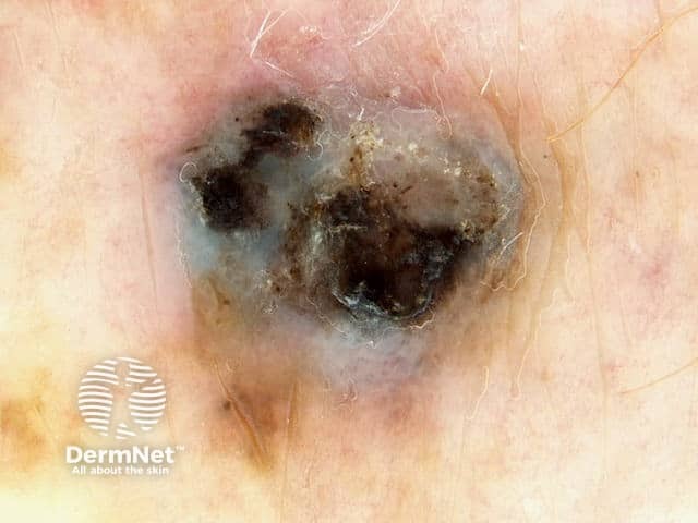
Right Lower Leg
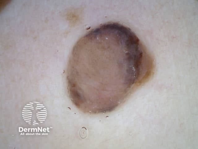
Left Posterior Shoulder
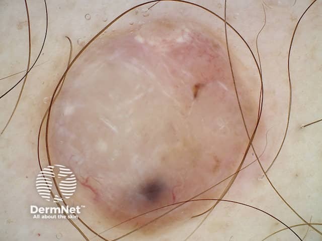
Nodular melanoma, Breslow 7.2 mm, polarised dermoscopy view
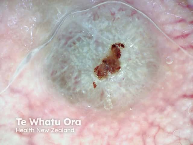
Nodular melanoma, Breslow 6.8 mm

Dermoscopy of nodular melanoma Breslow 4 mm

An ulcerated nodular lesion with polymorphous vessels and structureless area - a nodular malignant melanoma (NM-patient1)
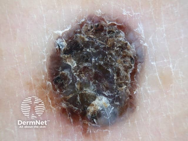
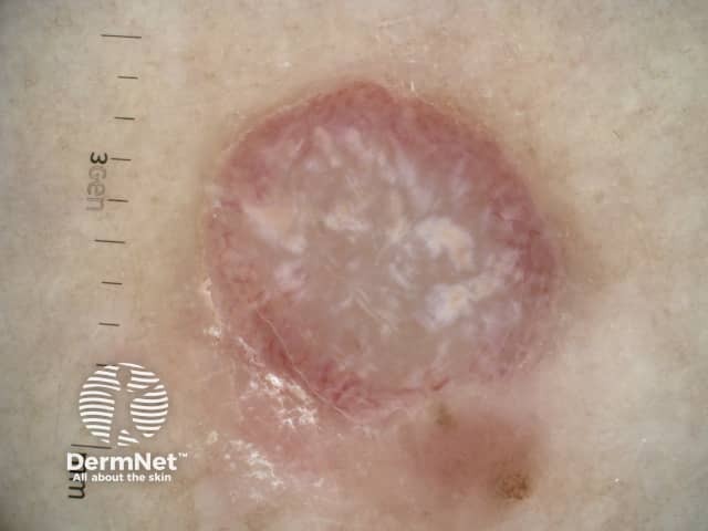
Right Lower Leg Outer