DermNet provides Google Translate, a free machine translation service. Note that this may not provide an exact translation in all languages

Translate

Quiz
Pigmented lesions – 10 cases

There are 10 pigmented lesions to diagnose in this quiz

For each of the ten cases, study the image(s) and then answer the questions. You can click on the image to view a larger version if required.

Each case should take approximately 2 minutes to complete. There is a list of suggested further reading material at the end of the quiz.

When you finish the quiz, you can download a certificate.
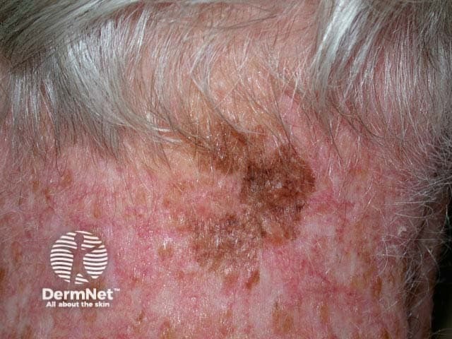
Case 3

This is a melanoma in situ. This means that the malignant cells are confined to the tissue of origin, the epidermis, and have not invaded through the basement membrane into the dermis.

It is not usually possible to be certain that a melanoma is in situ, although the clinical and dermoscopic appearance can be helpful. The pigmented lesion often appears to be a simple freckle at first. It may take several years to enlarge enough to come to the patient's attention. Look for irregularity in the shape of the lesion and variability in colour. If in doubt, cut it out!

Mark question

Sign up to the newsletter