Introduction Histology Special studies Differential diagnoses
Aggressive digital papillary adenocarcinoma are acral tumours, which have a broad morphologic spectrum and may cause diagnostic confusion. Previously, tumours would be divided into adenoma and adenocarcinoma based on atypia, but this has been revised: even bland tumours may behave in a malignant fashion.
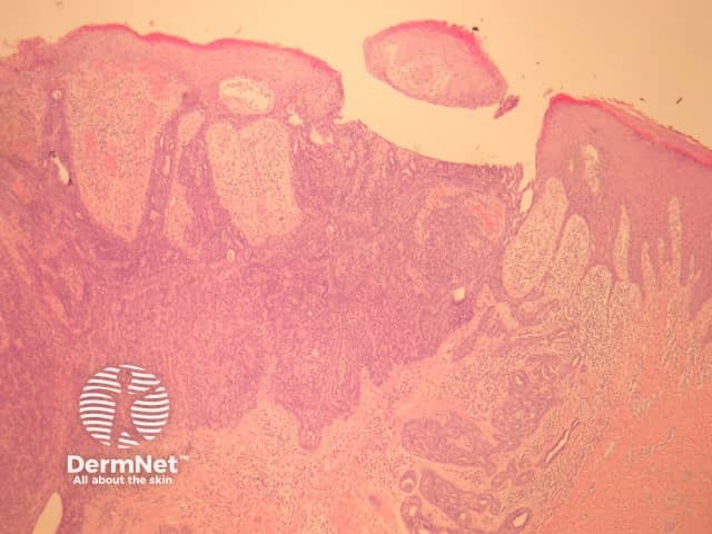
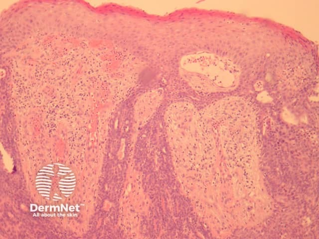
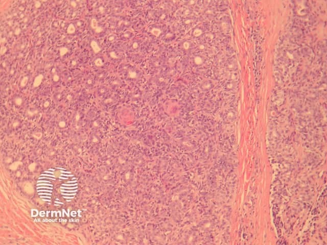
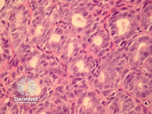
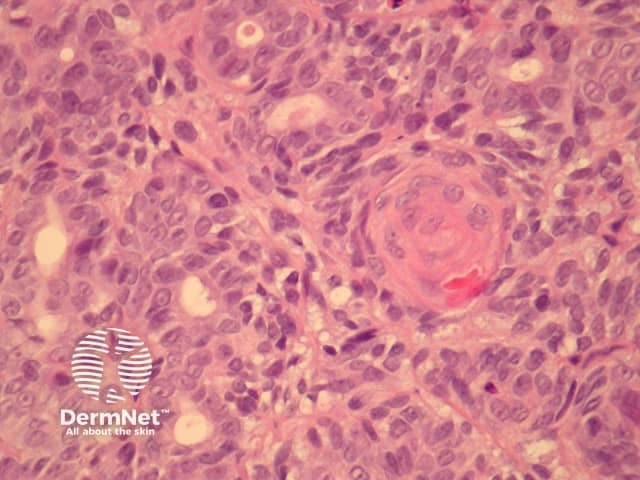
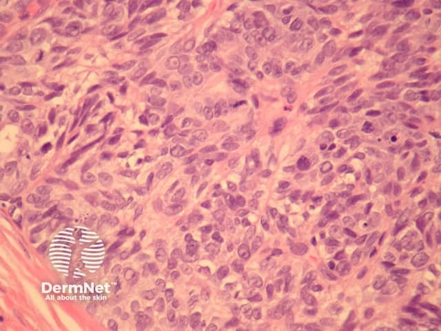
In aggressive digital papillary adenocarcinoma, sections show a dermally based tumour, which may connect with the overlying epidermis (figure 1). The tumour may be cystic, papillary, ductal or solid. The example illustrated here has a predominantly solid and ductal morphology with only focal areas of papillary formation. The cells are basaloid, show enlarged atypical nuclei, large nucleoli and increased mitoses (figures 1-6). Squamous metaplasia within the tumour may be seen (figure 5).
None are generally needed. In cases which are encountered in lymph nodes immunohistochemical studies to rule out metastasis from breast, thyroid or gastrointestinal sites may be needed.
Metastatic adenocarcinoma – Clinical history and immunohistochemical studies may be needed to rule out a dermal or nodal metastasis from another site.
Benign adnexal tumour – Some of these tumours can be deceptively bland. The acral site, infiltration of deep structures and epidermis and growth pattern helps in arriving at the correct (malignant) diagnosis.

