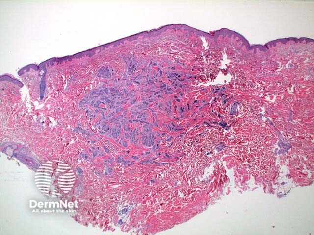
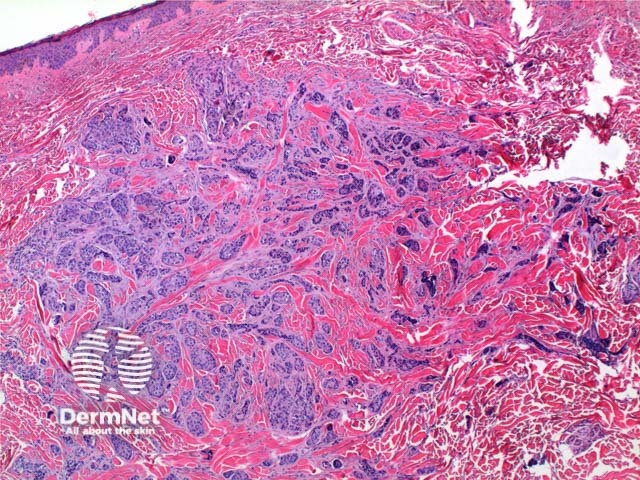
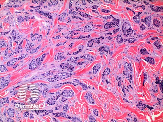
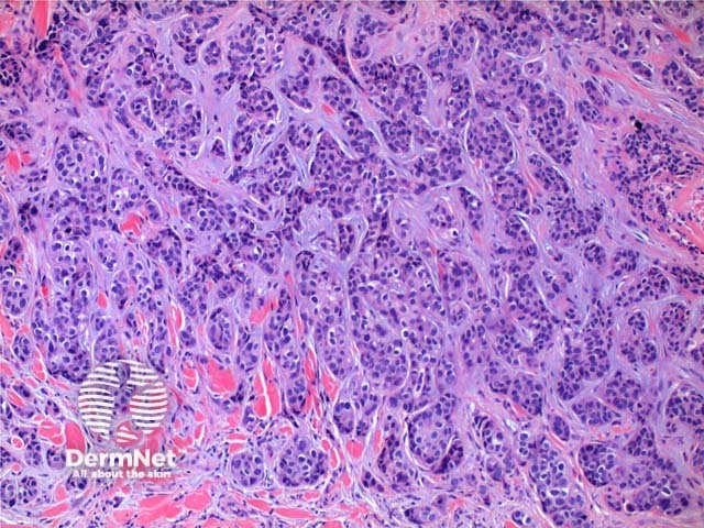
The histology of metastatic adenocarcinoma may show a number of patterns. Low power view frequently shows a poorly circumscribed infiltrating tumour centred on the dermis (Figure 1). Cords and nodules of atypical epithelial cells can be seen dissecting between collagen bundles (Figure 2). These may show evidence of duct or gland formation (Figure 3), and may be set in a mucinous stroma (Figure 4). Vascular and lymphatic permeation may be evident in the telangiectoides and erysipeloides variants of breast metastases.
While there is no substitute for clinical correlation and staging investigations, immunohistochemistry can provide clues to the site of origin, and help discriminate from primary cutaneous adnexal tumours. While never entirely specific, general rules are outlined below.

