Introduction Histology Special studies Differential diagnoses
Angioleiomyoma is an uncommon smooth muscle tumour arising from the smooth muscle of the vessel wall.
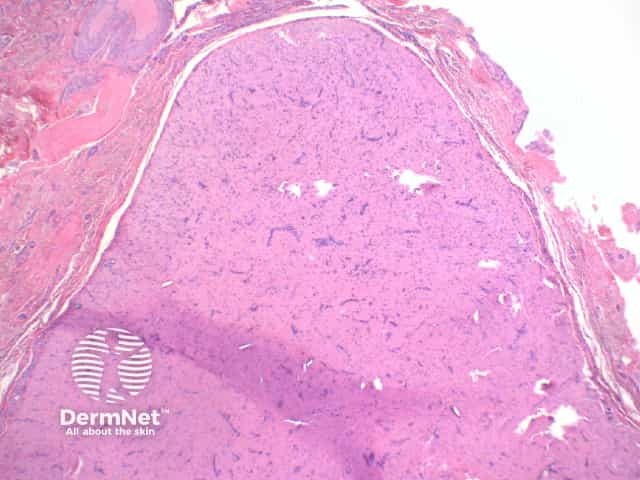
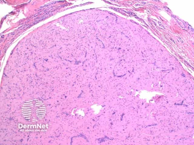
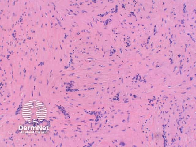
Scanning magnification view of angioleiomyoma shows a circumscribed tumour nodule arising in the dermis or subcutaneous tissue (figure 1). The tumour is comprised of densely packed interlacing bundles of smooth muscle which surround small compressed vascular channels (figures 2, 3).
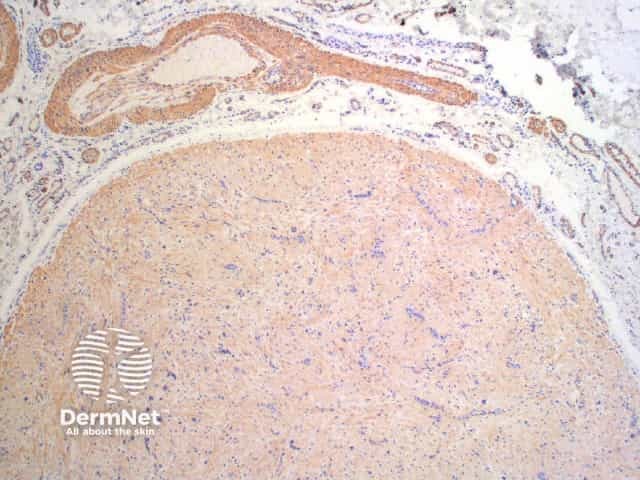
Immunostaining shows positive staining with muscle markers, Smooth muscle actin (SMA) and Desmin (figure 4 – SMA).
Myopericytoma: While this entity is also a circumscribed nodule which stains positively for muscle markers, it is comprised of perivascular concentrically arranged plump spindle to oval cells (glomoid-like) with eosinophilic cytoplasm.

