

Angiolymphoid hyperplasia with eosinophilia pathology — extra information
Introduction Histology Special studies Differential diagnoses
Introduction
Angiolymphoid hyperplasia with eosinophilia (or eosinophils) is also known as epithelioid or histiocytoid haemangioma (See also Epithelioid haemangioma pathology). It usually effects young adults and presents as clustered small, translucent nodules on the head and neck.
Histology of angiolymphoid hyperplasia with eosinophils
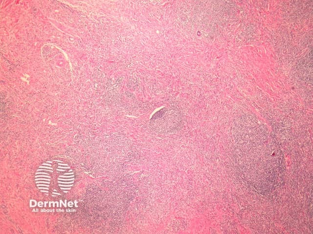
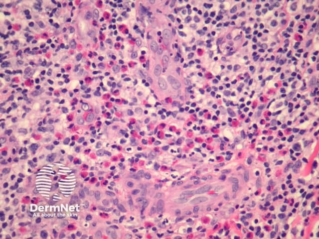
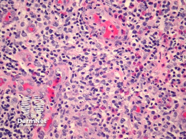
Sections show an inflamed dermis which shows increased vascularity (figure 1). Higher power examination reveals the endothelial cells lining the vessels are enlarged and epithelioid (figure 2, 3). The infiltrate is composed of lymphocytes which form follicles and numerous eosinophils (figures 2, 3).
Special studies of angiolymphoid hyperplasia with eosinophils
None are generally needed.
Differential diagnosis of angiolymphoid hyperplasia with eosinophils
Kimura disease – Can closely resemble angiolymphoid hyperplasia with eosinophilia. Eosinophilic abscess, eosinophils infiltrating germinal centers and dermal sclerosis are more common in Kimura disease. The vessels of Kimura disease generally lack an epithelioid endothelial lining.
References
- Pathology of the Skin (Fourth edition, 2012). McKee PH, J. Calonje JE, Granter SR