Introduction
Demographics
Causes
Clinical features
Complications
Diagnosis
Differential diagnoses
Treatment
Outcome
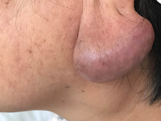
Kimura disease (KD) is a rare, chronic, inflammatory disorder.
It is characterised by subdermal lymphoid masses that are typically found in the head and neck region and/or cervical lymphadenopathy.
Kimura disease primarily occurs in young males of East Asian heritage. However, there have been case reports of Kimura disease in people from other ethnic backgrounds (eg, Hispanic, Middle Eastern, African) and other age groups (eg, young children).
The peak age of incidence is thought to be in the second and third decade of life.
The exact cause of Kimura disease is not known. Potential causes that have been proposed include infection, trauma, autoimmune processes, and allergic reactions.
Kimura disease presents as painless nodules in subcutaneous tissue, typically in the head and neck region, which normally have a diameter from 1–7 centimetres. Associated regional lymphadenopathy is common.
Other sites that may be involved include the oral cavity, orbit, nasal sinuses, the parotid gland, the axilla, and the groin. Atypical presentations include eczema, generalised pruritus, and prurigo nodularis. Patients do not usually present with systemic symptoms.
In conjunction with a clinical history and examination, a biopsy of the subcutaneous masses and/or enlarged lymph nodes can help with the diagnosis of Kimura disease.
Histopathological components of Kimura disease include:
Blood tests typically show eosinophilia and raised serum immunoglobulin E (IgE) levels. Other laboratory findings can include raised serum creatinine and albuminuria in the context of renal impairment and nephrotic syndrome.
Imaging studies are often non-specific but may help assess the extent and progression of Kimura disease, and any lymph node involvement.
Recurrence is common regardless of what treatment option is chosen. There have been no reported cases of malignant transformation. Patients who develop renal complications have varied outcomes dependent on the type and severity of nephropathy. Patients should continue to be monitored for recurrence and potential renal involvement.

