Introduction Clinical features Dermoscopic features Differential diagnoses Histology
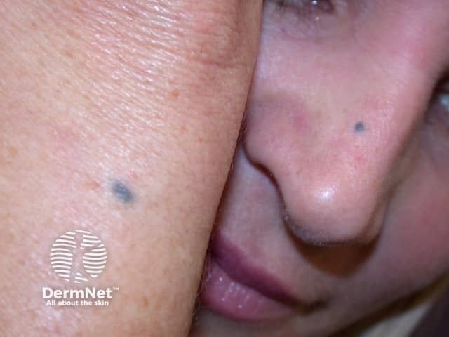
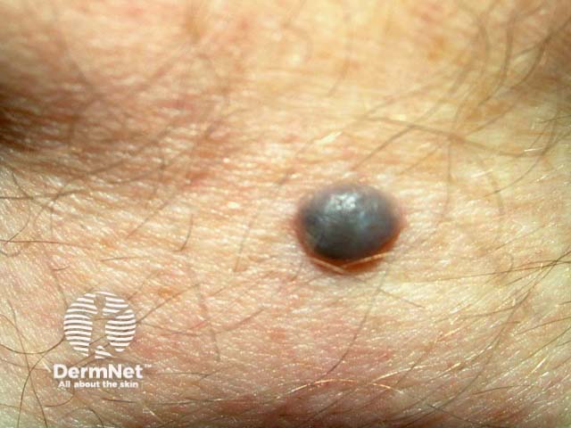
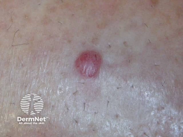
A blue naevus is a common type of melanocytic naevus in which the pigment is situated deep in the dermis. It is also known as a blue neuronaevus and a dermal melanocytoma.
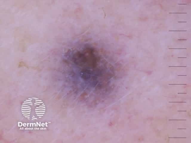
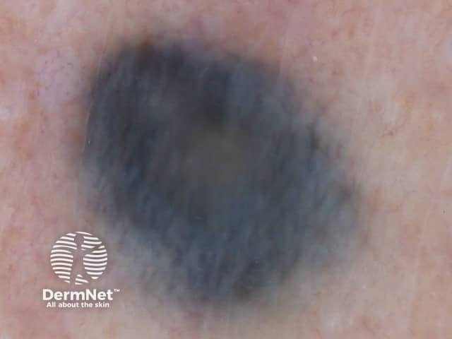
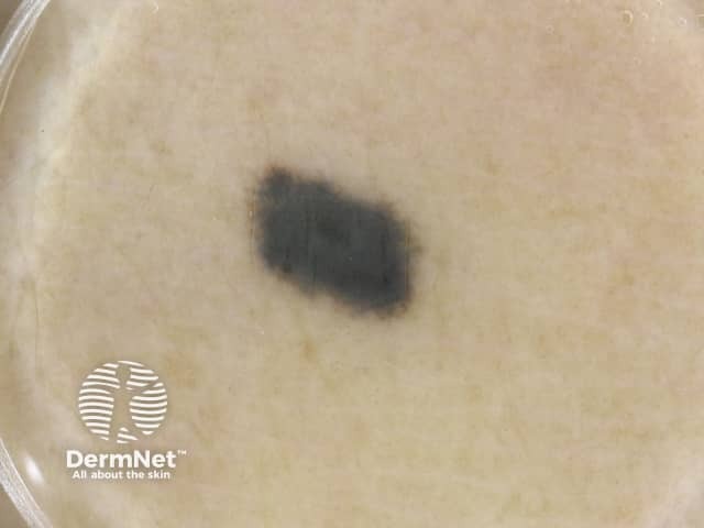
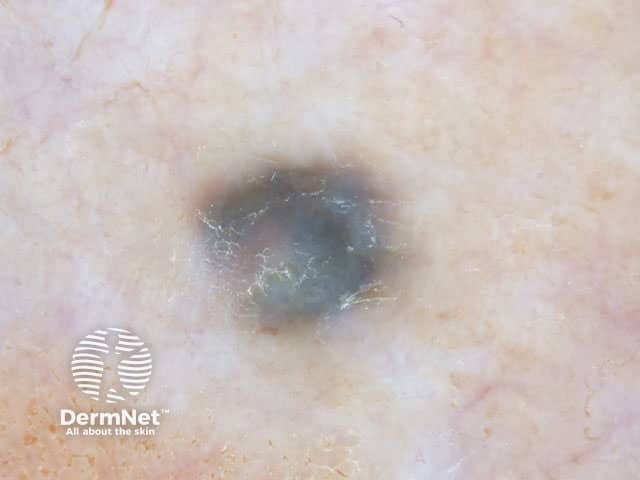
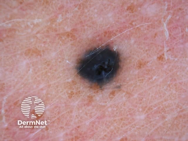
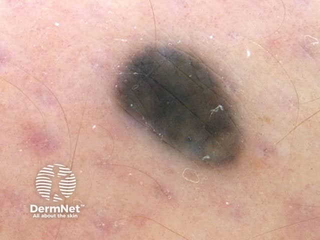
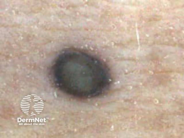
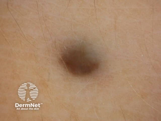
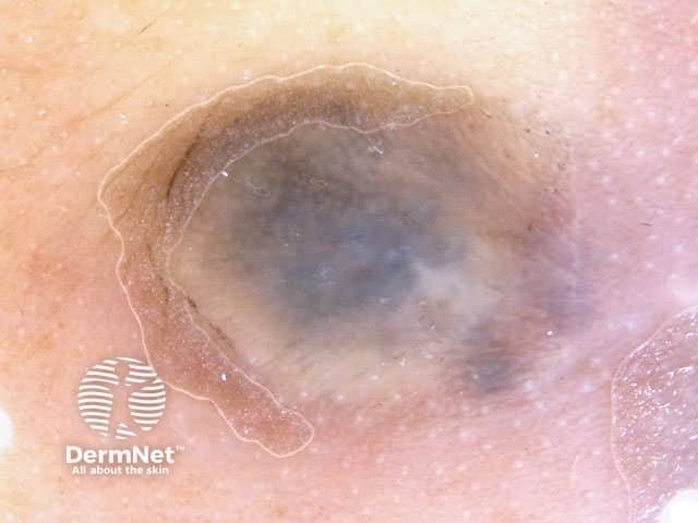
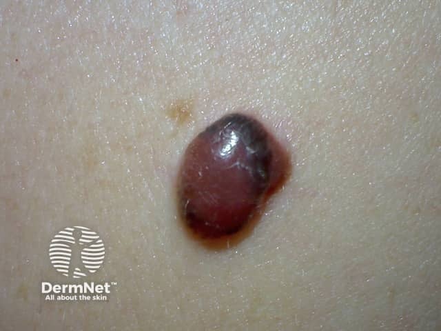
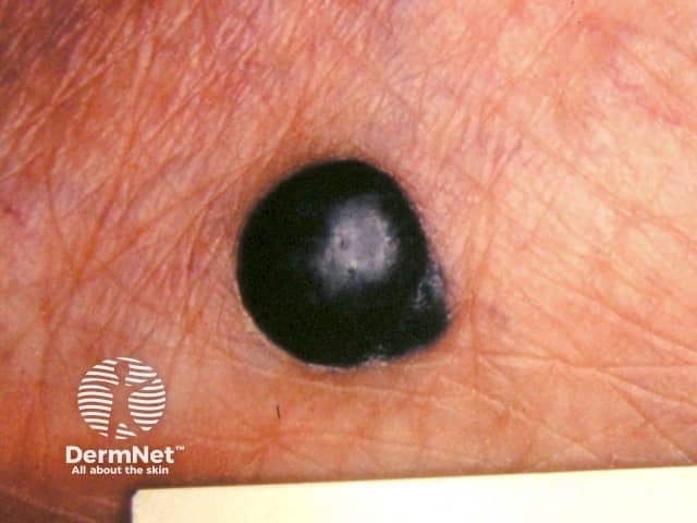
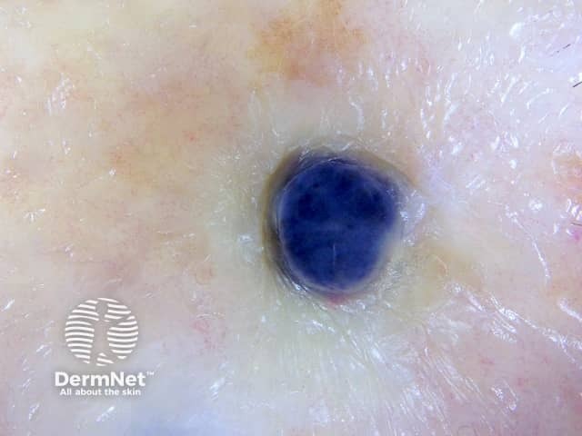
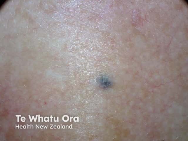
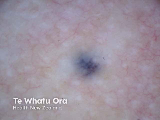
A blue naevus is a well-circumscribed round or oval macule, papule or nodule of a uniform steel blue colour. The border fades gradually into the surrounding skin.
In adults, the lesion is often long-standing. The most common age of onset is late childhood or adolescence.
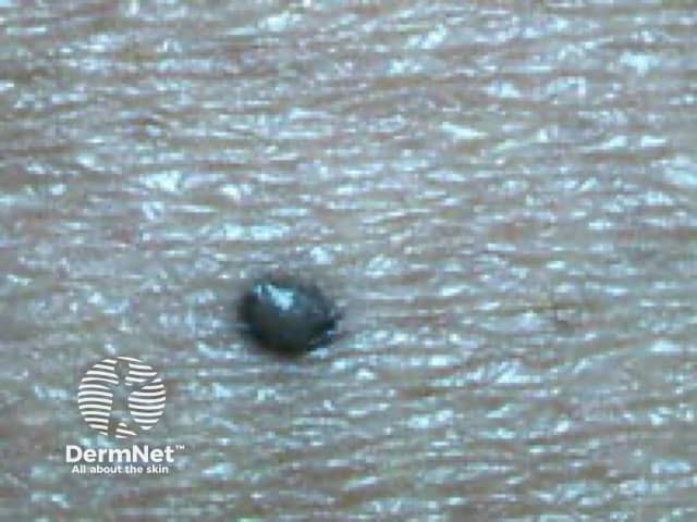
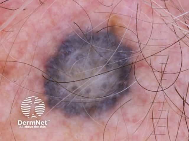
The dermoscopic hallmark of a blue naevus is homogeneous blue pigmentation. In some exceptional cases, they may exhibit blue globules and dots [1].
Dermoscopic features of a blue naevus are:
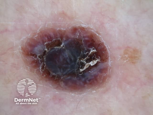
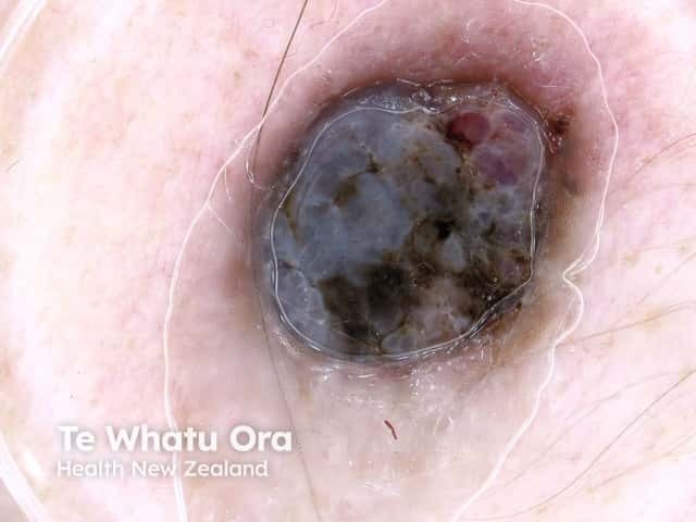
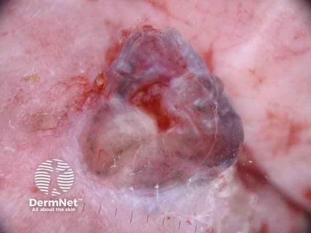
The presence of any of the following structures should warrant further investigations for melanoma: network, dots, clods, streaks, vessels, additional colours.
The primary concern of a lesion presenting as a blue naevus with unknown history is that it could be one of the following:

The histological explanation of a blue naevus is:

