Introduction Histology Special studies Differential diagnoses
Branchial cleft cysts are remnants of embryonic development and result from a failure of obliteration of one of the branchial clefts, which in fish develop into gills.
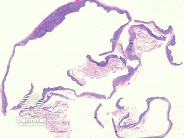
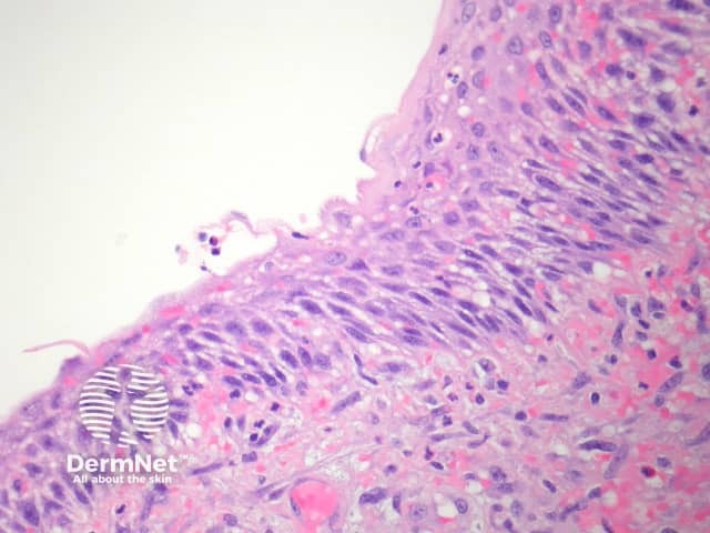
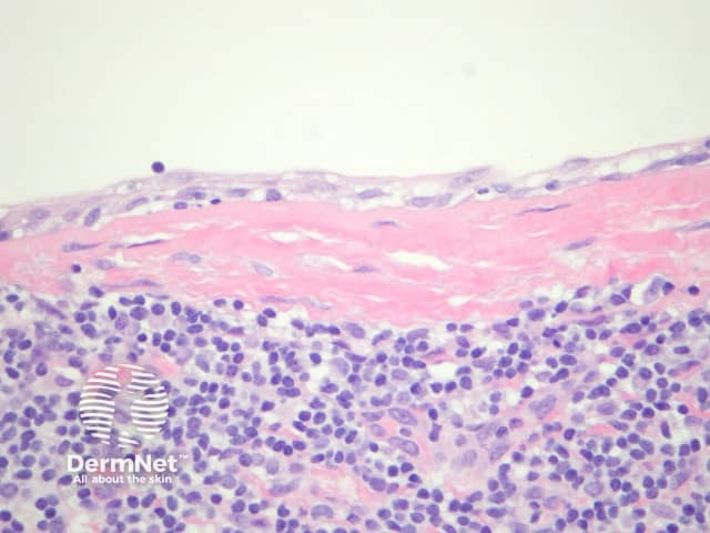
A branchial cleft cyst is often surrounded by lymphoid tissue (figure 1). The lining of the cyst is usually a stratified squamous epithelium (figure 2). The lining may also be a columnar ciliated epithelium. Often there are marked inflammatory changes and the epithelium overlying the lymphoid tissue is attenuated/absent (figure 3). Smooth muscle is rarely seen in the wall. Mucous glands and cartilage may also sometimes be seen in the wall.
None are needed.
Cutaneous ciliated cyst – Smooth muscle, mucous glands and cartilage are not seen in the walls of cutaneous ciliated cysts.
Metastatic squamous cell carcinoma – Squamous cell carcinoma metastatic to neck lymph nodes can be extremely difficult to distinguish from branchial cleft cysts, particularly when the metastatic tumour is cystic.
Bronchogenic cyst – Usually arise in the midline, almost always have smooth muscle or glands in the wall and are generally not associated with lymphoid tissue.

