Cutaneous plasmacytoma is a rare (< 1%) manifestation of multiple myeloma or plasma cell leukaemia. It disproportionately affects IgA and IgD proliferations and is associated with a worsened prognosis.
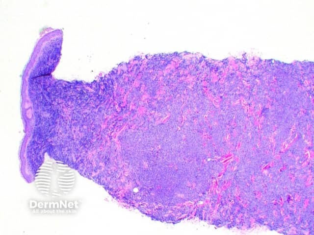
There is a dense cellular infiltrate (Figure 1) filling the biopsy specimen. This is comprised of a monotonous population of plasma cells (figure 2).
There are histologic grading criteria described by Bartl et al in 1987. Cases can be classified as well differentiated (grade I), moderately differentiated (grade II), or poorly differentiated (grade III) based on plasma cell morphology.

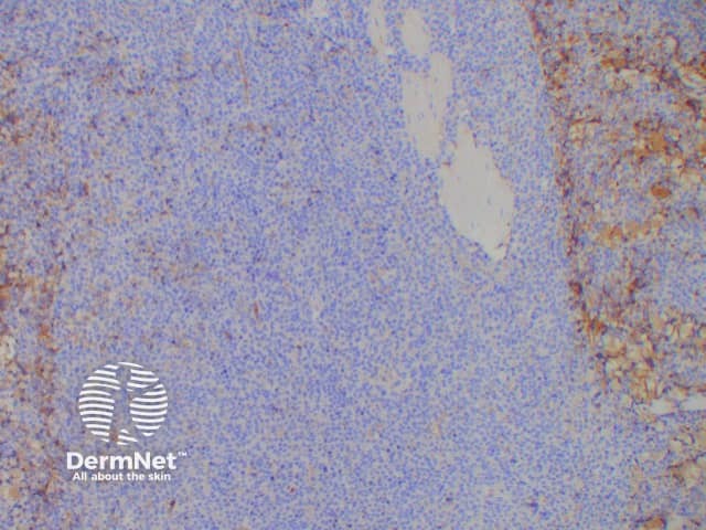
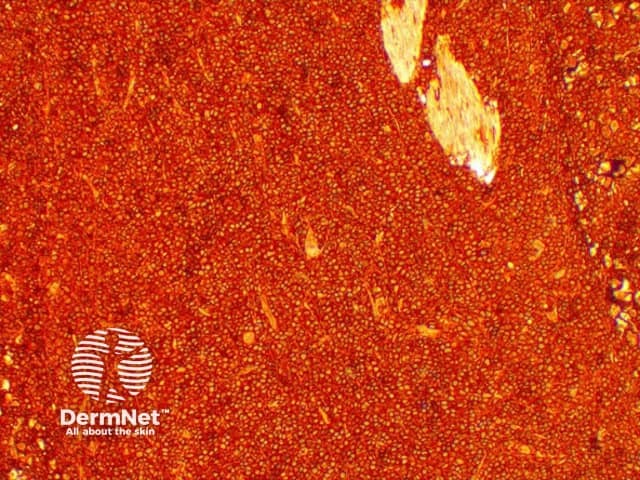
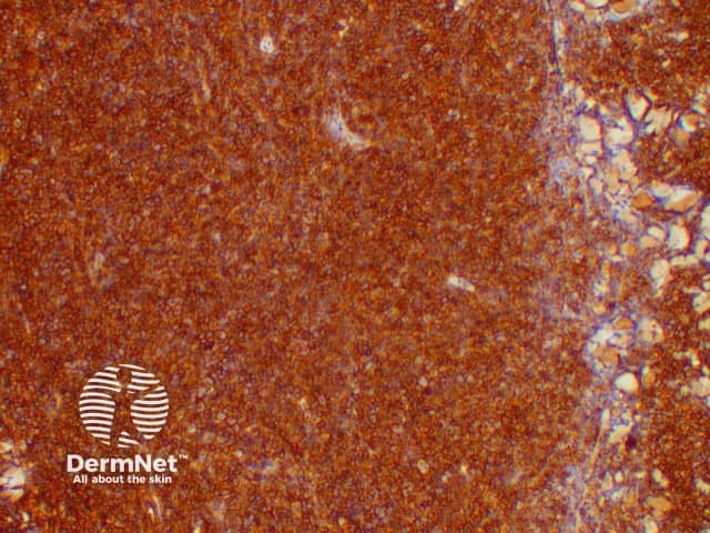
The plasma cells show a monoclonal profile confirmed by increased kappa or lambda staining on immunohistochemistry (figures 3, 4) or FISH. Plasma cells are also highlighted by CD138 immunostaining (figure 5).