

Darier disease pathology — extra information
Introduction Histology Histological variants Differential diagnoses
Introduction
Darier disease belongs to the group of acantholytic dyskeratoses, characterised by the presence of suprabasal separation due to the process of acantholysis with focal dyskeratosis of the keratinocytes.
Histology of Darier disease
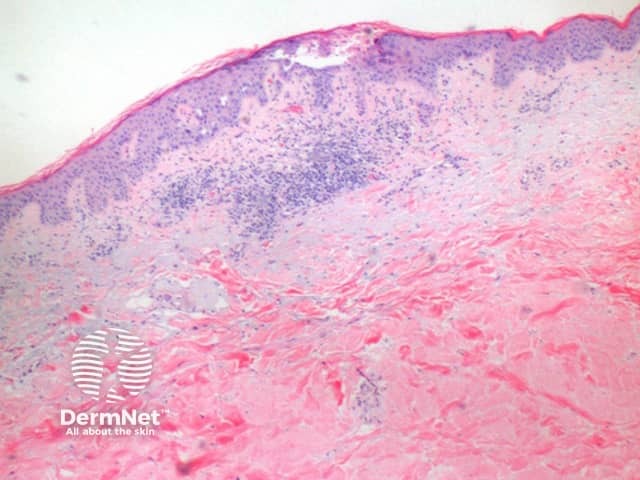
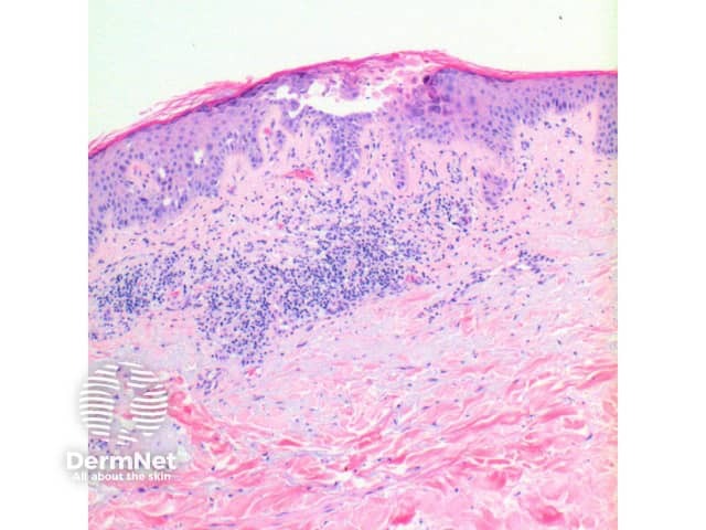
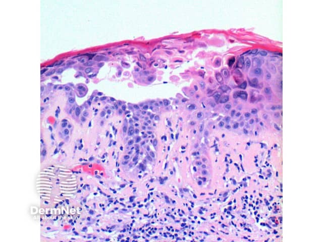
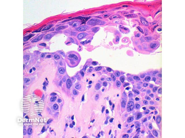
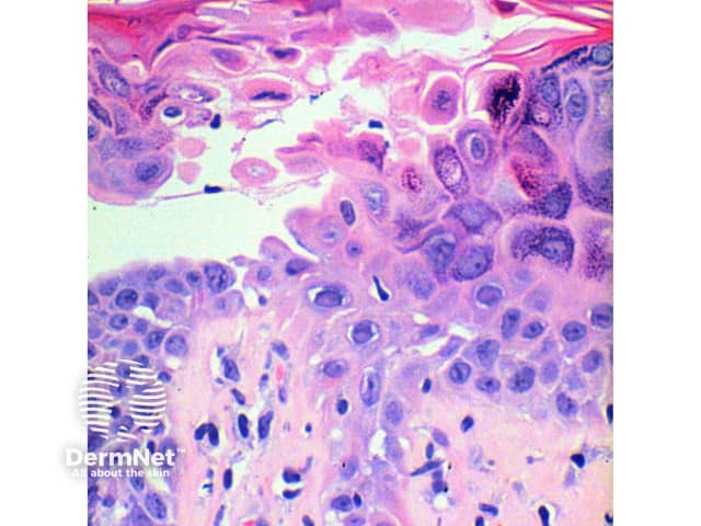
The scanning power view of the histology of Darier disease is of an epidermal and superficial dermal inflammatory process (Figure 1). Intraepidermal separation may be visible at this power, which on closer inspection is seen as suprabasal acantholysis (Figures 2 and 3). Acantholysis can be seen at all levels within the epidermis. Dyskeratosis of the keratinocytes is seen, with two notable changes described. Corps ronds refer to cells with small pyknotic nuclei, a perinuclear clear halo and eosinophilic cytoplasm (Figures 4 and 5).
Grains are compressed cells with elongated nuclei seen in the stratum corneum and granular layer (Figures 4 and 5).
The overlying epidermis exhibits compact orthokeratosis, which can form a prominent focal plug.
In the superficial dermis is a mild predominantly perivascular lymphocytic infiltrate (Figure 2). Eosinophils are rarely seen.
Histological variants of Darier disease
Comedonal Darier disease: in this variant the foci of acantholytic dyskeratosis have more pronounced keratotic plugs and deeper involved hyperplastic extensions of the epidermis into the superficial dermis.
Bullous Darier disease: here the difference is purely in the size of the acantholytic clefts, which form lacunae with scattered acantholytic cells within.
Acrokeratosis verruciformis of Hopf: While debated whether this is a component of Darier disease or a distinct condition, the changes are briefly mentioned here, given the clinical association. The lesions exhibit church spire papillomatosis with hyperkeratosis and epidermal acanthosis. If heavily sectioned, small foci of acantholytic dyskeratosis may be seen.
Differential diagnosis of Darier disease
Hailey-Hailey disease: the key difference here is the breadth of involvement of the epidermis by the acantholysis. There are more pronounced areas of partially and completely acantholytic cells.
Transient acantholytic dermatosis/Grover disease: while the Darier-like variant of Grover disease may show near-identical features, the presence of smaller involved foci, and an inflammatory infiltrate containing eosinophils serve as clues to this diagnosis.
References
- Skin Pathology (2nd edition, 2002). Weedon D
- Pathology of the Skin (3rd edition, 2005). McKee PH, J. Calonje JE, Granter SR
- Telfer NR, Burge SM, Ryan TJ. Vesiculobullous Darier's disease. Br J Dermatol. 1990; 122:831–4
On DermNet