

Eccrine papillary adenoma pathology — extra information
Eccrine papillary adenomas are benign sweat gland tumours that have been described in wide range of anatomic sites and age groups.
Histology of eccrine papillary adenoma
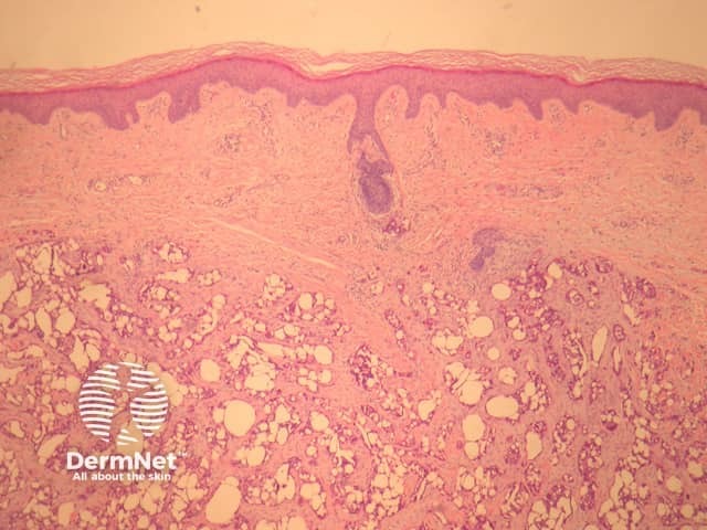
In eccrine papillary adenoma, sections show a well-circumscribed dermal tumour set in a sclerotic stroma (figure 1).
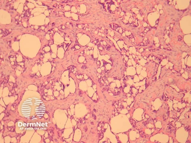
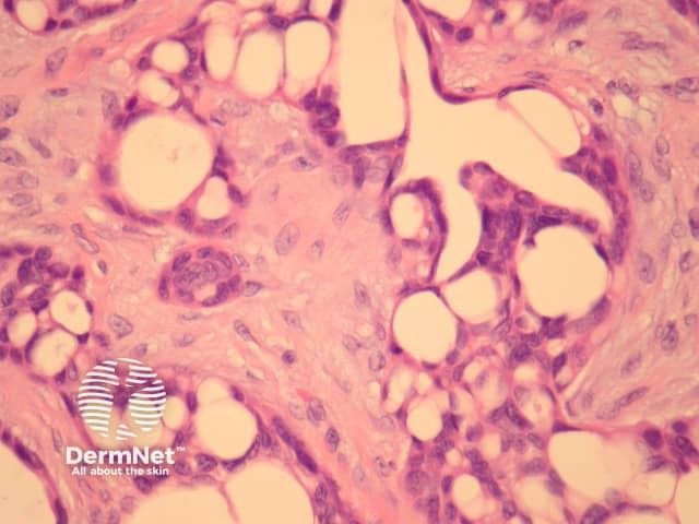
The tumour forms tubular and tubulo-papillary structures (figures 2, 3). The ducts are mainly lined by 2 or more cell layers of bland epithelial cells with limited mitotic activity. Some cells have an eosinophilic cytoplasm.
Special studies of eccrine papillary adenoma
None are generally needed.
Differential diagnosis of eccrine papillary adenoma
Metastatic tumour – These usually have an infiltrative growth pattern with more nuclear atypia. Clinical history and immunohistochemical studies can be helpful for unusual cases.
Aggressive digital papillary adenocarcinoma – These often have solid areas, arise on acral sites and generally show more atypia.
Microcystic adnexal carcinoma – These have an infiltrative growth pattern which may involve nerves and areas squamous differentiation.
References
- Pathology of the Skin (Fourth edition, 2012). McKee PH, J. Calonje JE, Granter SR
On DermNet