

Epithelioid haemangioma pathology — extra information
Introduction Histology Special studies Differential diagnoses
Introduction
Epithelioid hemangioma is a benign vascular lesion of the skin or deeper structures (bone). It has many similarities to angiolymphoid hyperplasia with eosinophilia and some authors believe they are the same entity. Although eosinophils are often rich in the lesion, peripheral eosinophilia is not a feature.
Histology of epithelioid haemangioma
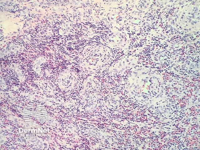
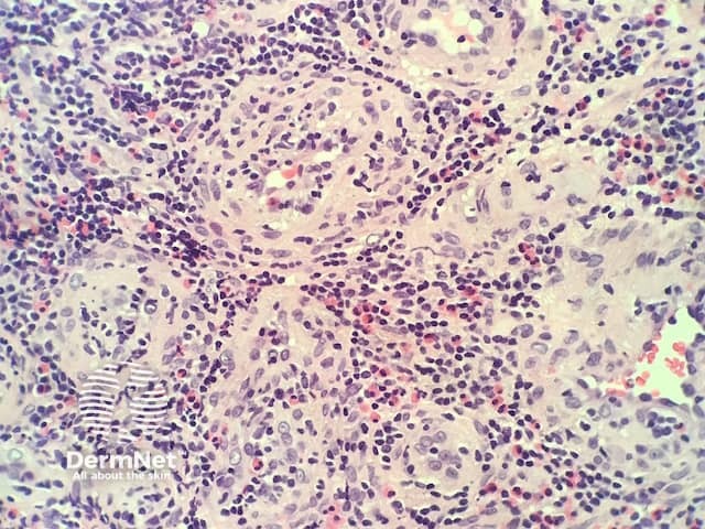
In epithelioid haemangioma, histopathologically there is a proliferation of blood vessels with epithelioid endothelial cells (figures 1,2). The endothelial cells are plump and have abundant eosinophilic cytoplasm sometimes resembling histiocytes (best seen in figure 2). As the endothelial cells are so plump, sometimes it is difficult to appreciate vascular spaces and the aggregates may resemble granulomas. Accompanying the vascular proliferation are collections of lymphocytes an numerous eosinophils (figures 1,2).
Special studies for epithelioid haemangioma
None are generally needed. Immunohistochemical markers for CD31, CD34 can be helpful to highlight the endothelial cells and the overall architecture of the lesion
Differential diagnosis for elastofibroma
Angiolymphoid hyperplasia with eosinophilia (ALHE) — Some authorities believe these are the same entity. However, in contrast to ALHE, epithelioid haemangioma can involve any body site and involve the deep soft tissue and bone. Epithelioid haemangioma is usually an isolated lesion where ALHE often occurs in multiplicity.
Angiosarcoma: Angiosarcoma typically shows greater nuclear atypia and an invasive growth pattern
Kimura disease: usually Asians with elevated serum eosinophils and IgE, usually regional lymphadenopathy
References
- Epithelioid hemangioma — PathologyOutlines.com
On DermNet
- Angiolymphoid hyperplasia with eosinophilia pathology
- Dermatopathology glossary
- Dermatopathology index