

Facial rashes — extra information
Categories:
ADVERTISEMENT
Erosions and crusting Dry or scaly rash Papulopustular rash Erythema Brown macules and patches Pale or white macules and papules Skin lesions
Facial rashes
Patients often present with quite mild signs when they have a facial lesion or rash — due to embarrassment — and the diagnosis may be tricky.
Significant itch suggests atopic dermatitis or contact dermatitis.
Face: erosions/crusting
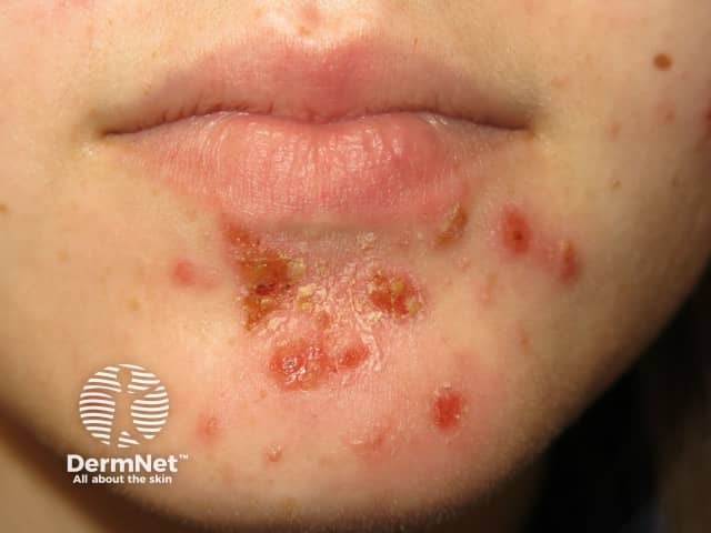
Herpes simplex
- Monomorphic clustered vesicles or crusted papules
- Often locally recurrent in the same site
- Swabs: Herpes simplex
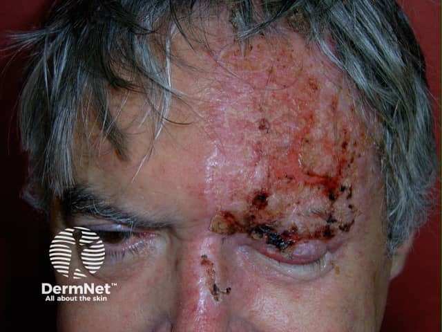
Herpes zoster
- Acute dermatomal eruption
- Painful: pain may precede the rash
- Erythema may precede vesicles
- Swabs: Herpes zoster
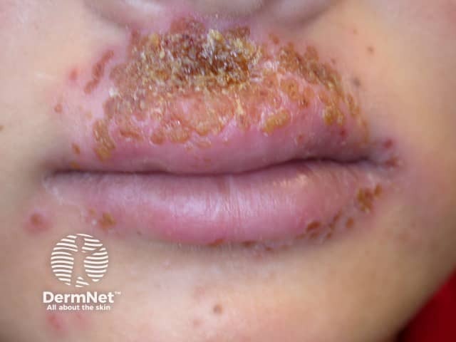
Impetigo
- Irregular enlarging plaque
- Honey-coloured crusts
- Swabs for impetigo: Staphylococcus aureus +/- Streptococcus pyogenes
Dry or scaly rash
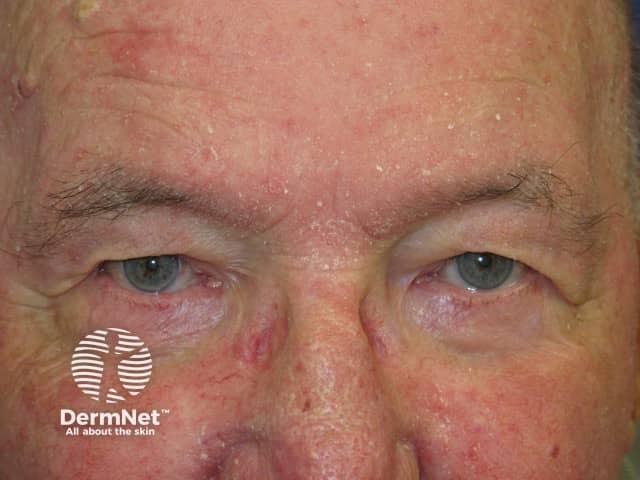
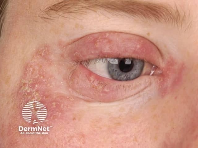
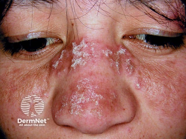
Seborrhoeic dermatitis
- Seborrhoeic dermatitis often also affects scalp, retroauricular sites, ears
- Hairline, eyebrows, medial cheeks, nasolabial folds, chin creases
- Scaly blepharitis
- Poorly defined, variable white or yellowish flaking
- May have erythematous patches or thin plaques
- Follicular prominence or follicular digitate keratoses
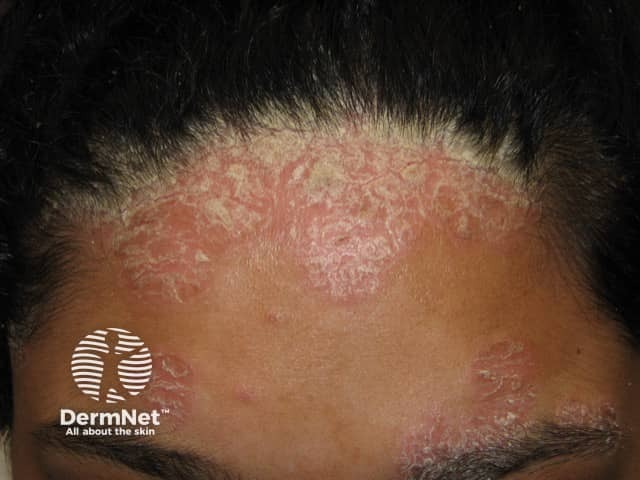
Psoriasis
- Psoriasis sites include eyelids, temples, retro- and pre-auricular skin and/or seborrhoeic dermatitis sites
- Also affects scalp, ears, elbows, knees, nails
- Well-demarcated erythematous plaques
- White scale
- More persistent than seborrhoeic dermatitis
Atopic eczema
- Atopic dermatitis often affects flexures: retroauricular, elbow and knee creases
- Symmetrical dermatitis of eyelids, perioral skin (up to lips)
- Intensely itchy
- Acute flare: oedema, erythema, crusting, fissuring
- Subacute: dryness, pinkness
- Chronic: dryness, lichenification, Dennie Morgan folds (2 creases in lower eyelids)
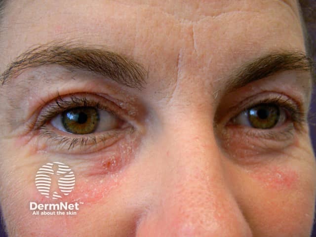
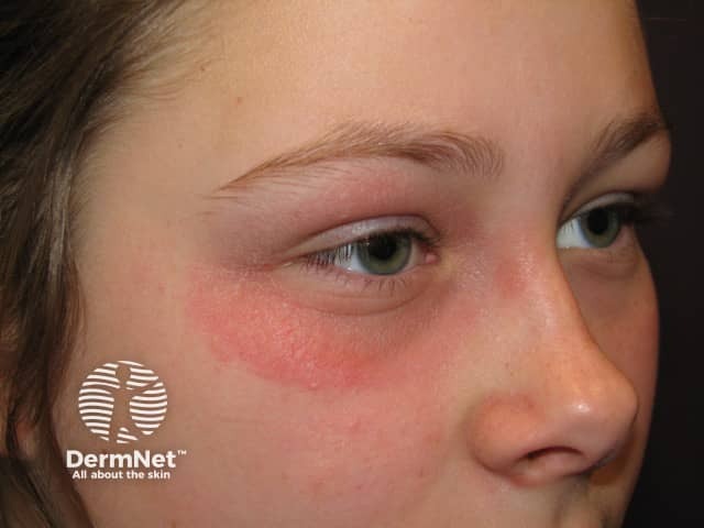
Contact eczema
- Acute, relapsing/intermittent or chronic presentation
- Irregular, variable, unilateral or asymmetrical dermatitis
- Sharp border if contact irritant dermatitis
- Patch tests positive if contact allergic dermatitis
Photosensitive dermatitis
- Exposed areas of face, arms, chest, legs
- Spares under hair, eyelids, creases
- Flares after exposure outdoors
- Can be drug-induced
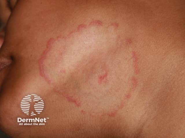
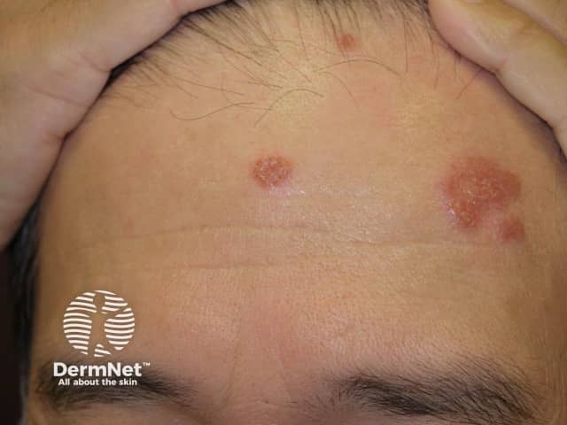
Tinea faciei
- Asymmetrical eruption
- Annular configuration is common
- Scaly edge
- Mycology positive
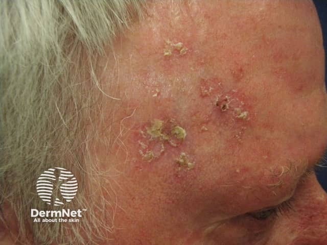
Actinic keratoses
- Located on sun-exposed sites of temples, forehead, nose, cheekbones, angle of jaw, upper lip, lower vermilion lip
- Actinic keratoses often involves persistent tender scaly papules, macules, plaques
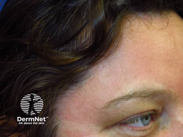
Cutaneous lupus erythematosus
- Nose, cheeks, ears, lips, scalp
- Circumscribed plaques with follicular prominence, scale
- Post-inflammatory pigmentation, atrophic scarring
- CBC, ANA, ENA often normal
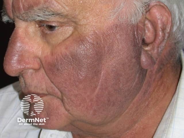
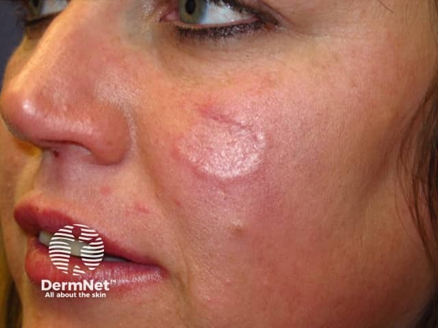
Lupus tumidus / Jessner lymphocytic infiltrate
- Cheeks, upper trunk
- Smooth surface to erythematous dermal plaques
Papulopustular rash
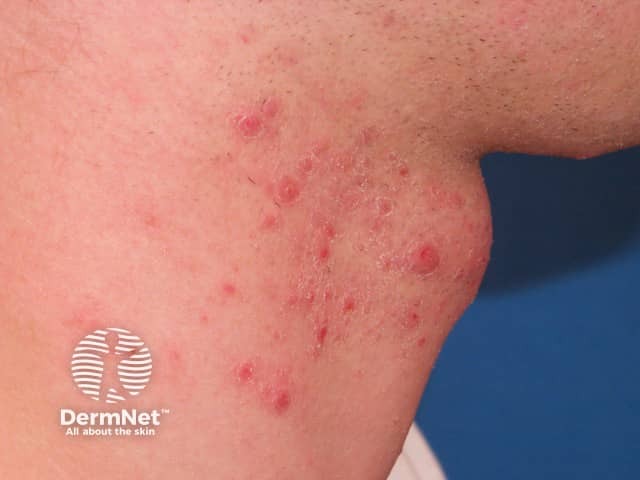
Acne
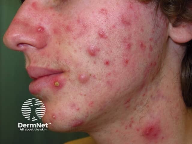
- Onset of acne often at puberty
- Usually, symmetrical forehead, chin, lateral face, nose
- Mixed inflammatory and non-inflammatory lesions
- Papules, pustules, nodules, comedones
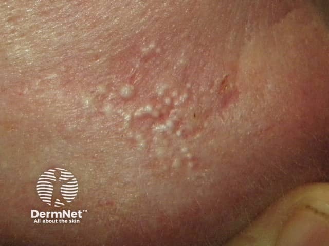
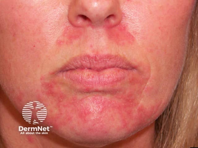
Perioral/periorificial dermatitis
- Usually adult females using face cream, often topical corticosteroid
- Often, asymmetrical first in perioral sites, later in perinasal and periocular sites
- Spares a centimetre of skin around vermilion of lips
- Grouped erythematous papules and pustules on erythematous patches, flaky surface
- Can occur in children
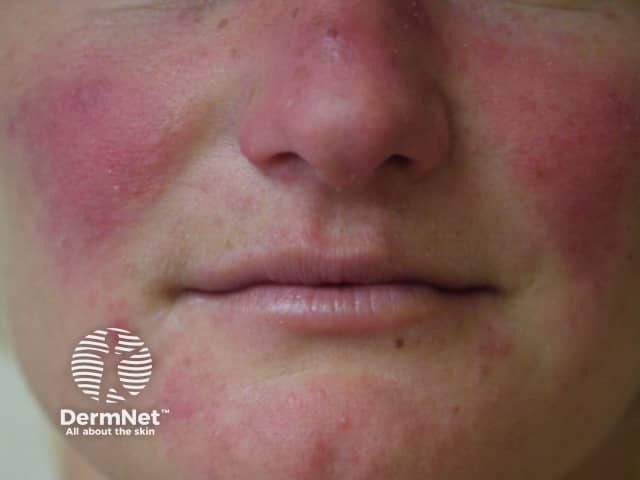
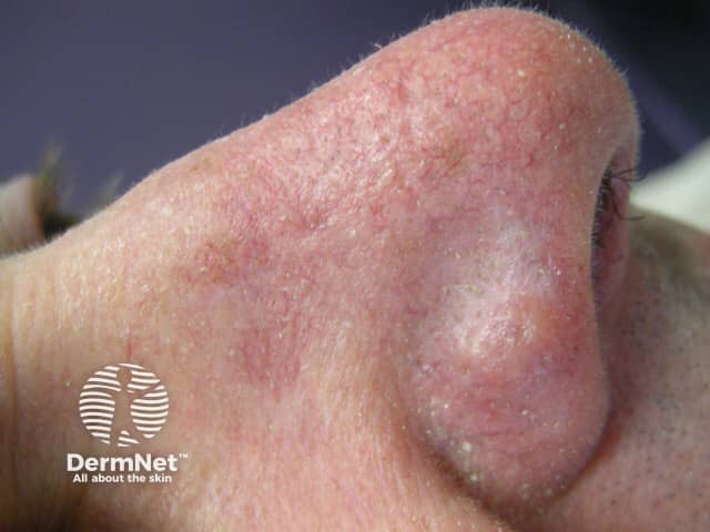
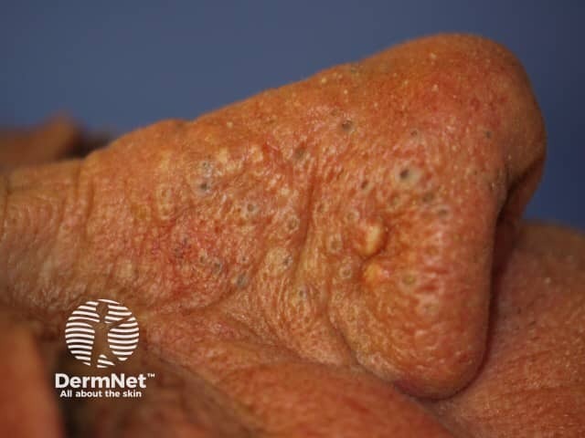
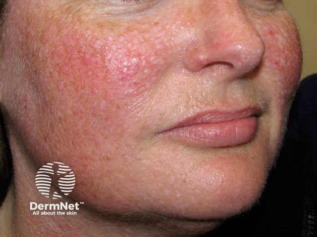
Rosacea
- Most prevalent in middle-aged adults
- Mid-facial: cheeks + nose, chin and forehead
- Erythema, flushing, papules, pustules, telangiectasia
- Rhinophyma causes enlargement of the nose in some patients
- Sensitive skin
- Lesions in rosacea can approach the lips
Pseudofolliculitis barbae
- Pseudofolliculitis barbae is most often associated with shaving
- Follicular papules, pustules, curled-in hair

Face: erythema
Erythema is less pronounced in dark skin
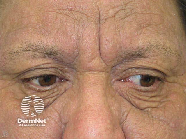
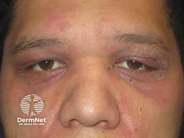
Dermatomyositis
- Violaceous eyelids — may be swollen
- Poikiloderma on the trunk and limbs
- Gottron papules on fingers
- May have muscle weakness
Flushing
- Intermittent redness when hot, embarrassed or with certain foods
- Often lifelong tendency
- Systemically well
- Associated with rosacea
Sunburn
- Sun-exposed site
- Spares eyelids, furrows, under the chin
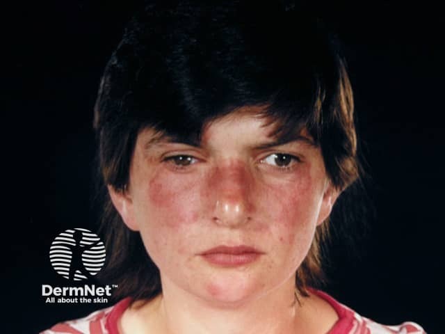
Systemic lupus erythematosus
- Butterfly erythematous rash
- Systemic symptoms: tiredness, lethargy, arthralgia
- Check CBC, ANA, ENA
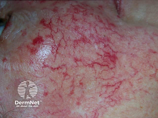
Telangiectasia
- May accompany flushing
- Vascular dilatation
- Various types
Face: brown macules/patches
Pigmentation is more pronounced in dark skin
Solar lentigines
- Sun-exposed sites
- Small to large freckles
- Well-demarcated flat or slightly scaly brown marks or thin plaques
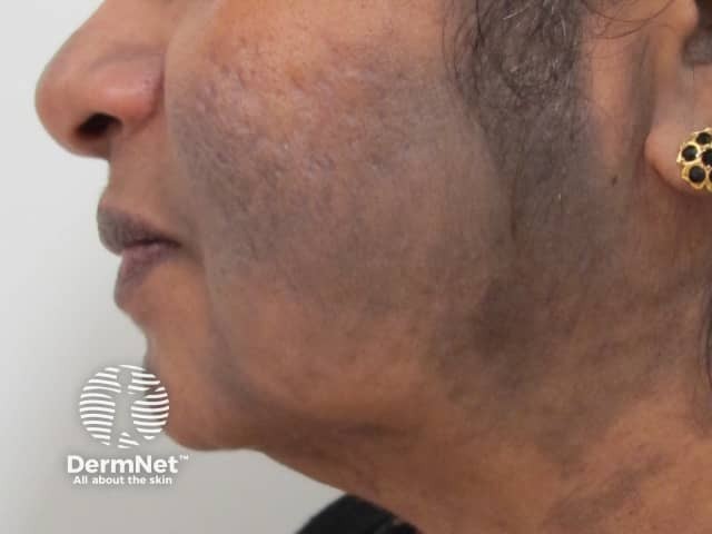
Erythema dyschromicum perstans
- Grey-brown discolouration
- Any distribution
- Distinct border, sometimes red at first
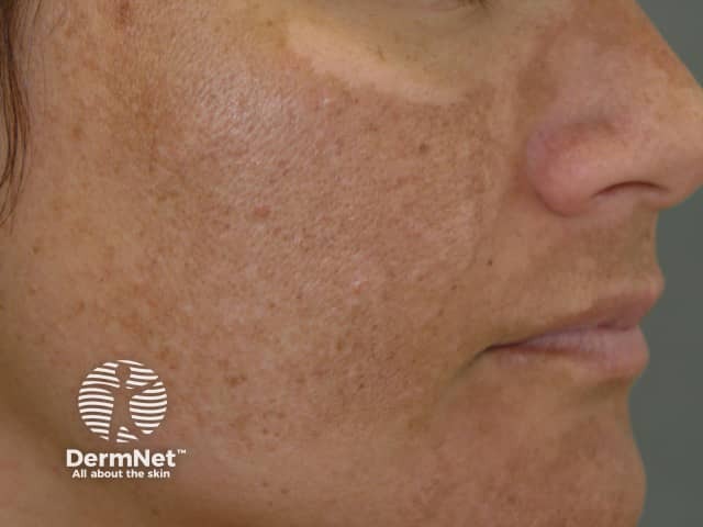
Melasma
- Usually adult female
- Centrofacial, malar and mandibular patterns
- Spares eyelids, rare below jawline
- Symmetrical pigmentation with ragged border
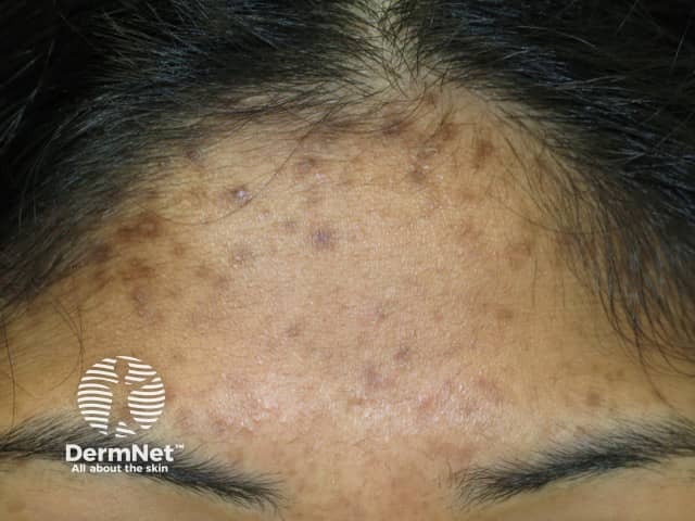
Post-inflammatory pigmentation
- Preceding eczema, psoriasis, acne etc
- Distribution depends on cause
Face: pale or white macules/patches
Guttate hypomelanosis
- More commonly observed on limbs
Pityriasis alba
- Young child
- Cheeks
- Hypopigmentation, light scale
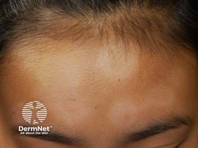
Post-inflammatory hypopigmentation
- Preceding eczema, psoriasis, acne etc
- Distribution depends on cause
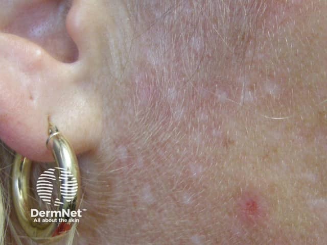
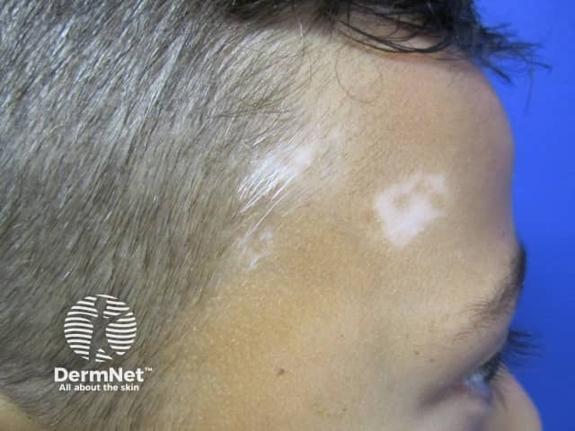
Vitiligo
- Most often periocular, perioral
- White, smooth surface
Skin lesions
Granuloma faciale
- Middle-aged adult
- Solitary thickened smooth, purplish-brown plaque or plaques
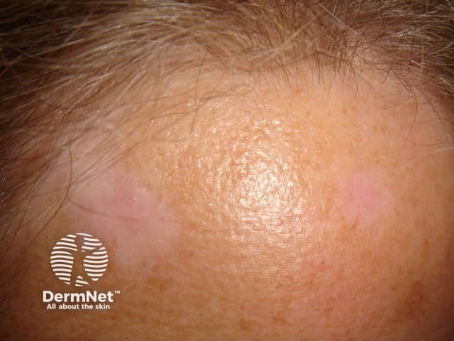
Sarcoidosis
- Yellowish-brown to mauve infiltrated plaque
- May arise within existing scar
- Lupus pernio affects nose and ears
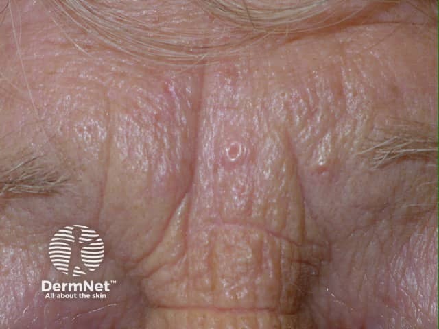
Sebaceous hyperplasia
- Mostly > 40 years
- Forehead, temples
- Yellowish papules with central follicular dimple
Solar comedones
- Smoker, sun damaged older patient
- Periocular, cheekbones, nose, neck
- Usually symmetrical
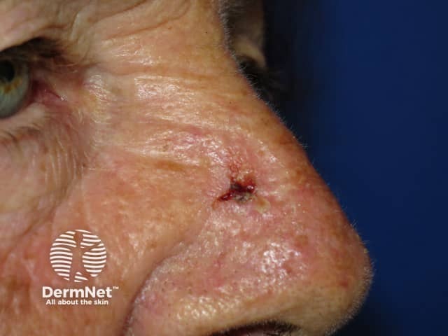
Basal cell carcinoma
- Slowly enlarging, destructive papule, nodule or plaque
- Early erosion, ulceration and bleeding
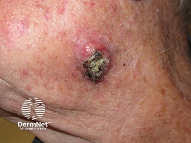
Squamous cell carcinoma
- Enlarging tender scaly or crusted nodule
Adnexal tumours
- Various types and syndromes
- Follicular or eccrine origin
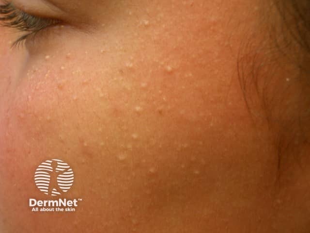
Milia
- Periorbital or cheeks
- Superficial firm small papules
- Scattered on forehead, cheeks
- Yellowish with central dell
ADVERTISEMENT
Other recommended articles
ADVERTISEMENT