Introduction Histology Histological variants Differential diagnoses
The finding of nodular elastosis with cysts and comedones associated with age, sun exposure and smoking, is known as the Favre Racouchot syndrome.
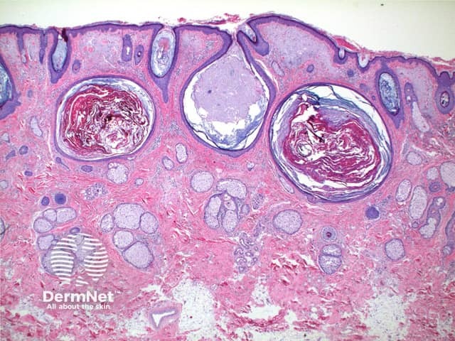
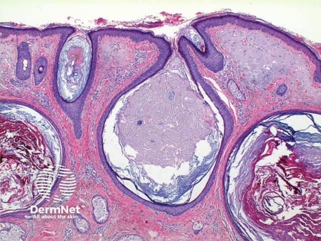
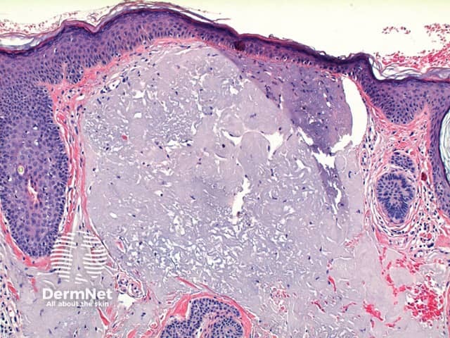
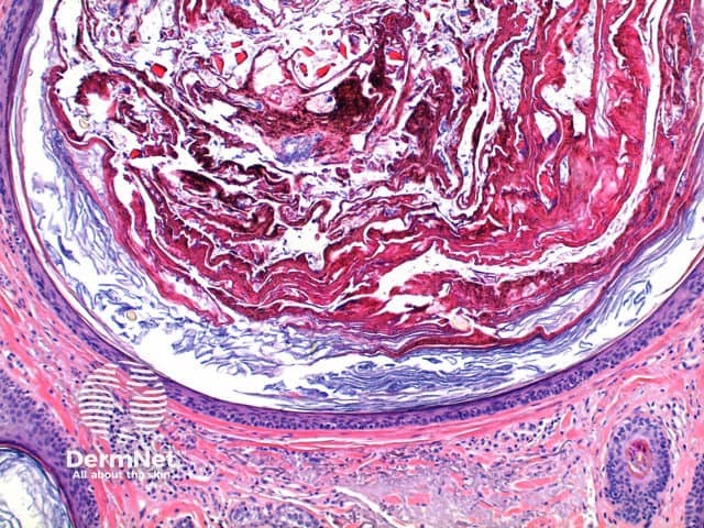
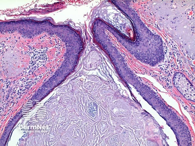
The low power view of Favre-Racouchot syndrome shows multiple comedones, dilated and plugged follicular infundibulae and epidermal cyst formation (Figures 1 and 2). This is set in a grossly solar damaged epidermis, which forms large nodules in the superficial dermis (Figure 3). The comedones are filled with compact keratinous material, while the epidermal cysts contain more loosely arranged laminated keratin (Figures 4 and 5).
Cases with cysts and comedones but without significant solar damage have sometimes been reported as Favre-Racouchot syndrome, with the suggestion that extensive sun exposure may not be essential pathogenetically in all cases.
Similar or identical changes may be seen in cases of chloracne or severe acne vulgaris in severely solar damaged skin, though less prominent cyst formation would be expected in these cases. Clinical correlation is essential here.

