Main menu
Common skin conditions

NEWS
Join DermNet PRO
Read more
Quick links
Follicular disorder Diagnosis and testing
Author: Dr Ben Tallon, Dermatologist/Dermatopathologist, Tauranga, New Zealand, 2011.
Introduction
Histology
Special stains
Differential diagnoses
Folliculitis keloidalis (also called folliculitis keloidalis nuchae) is best grouped as one of the neutrophilic scarring alopecias.
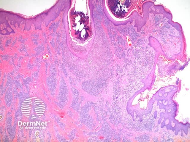
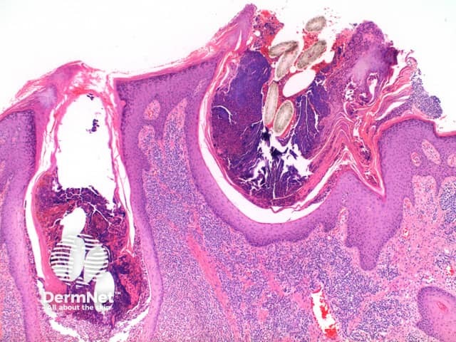
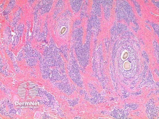
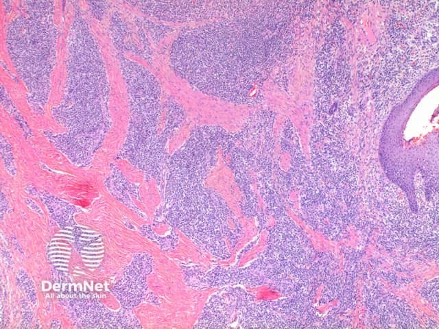
Low power view exhibits a dense superficial and deep inflammatory process with dermal scarring and follicular disruption (Figure 1). There may be variable degrees of overlying scale crust with tufted hair follicles evident as multiple hair shafts within widened follicular infundibulae (Figures 1 and 2). In the dermis are disrupted hair follicles with scattered naked hair shafts seen within a fibrotic dermis (Figures 2 and 3). There is a dense lymphoplasmacytic infiltrate with scattered neutrophils (Figure 4).

Figure 1

Figure 2

Figure 3

Figure 4
PAS staining should always be performed to exclude a fungal infection.
Folliculitis decalvans: While many features are shared, there is typically significantly less fibrosis in this condition. Clinical discrimination is reliable.
Hidradenitis suppurativa: While location and clinical details are discriminatory, this condition will show sinus tracts and large areas of abscess formation.
Deep infectious folliculitis: In these cases, the inflammatory infiltrate tends to form tightly around the involved follicle with less extensive follicular disruption or dermal scarring. Special stains are also of assistance here. In cases of doubt, culture studies should be recommended.