

Histology stains — extra information
Introduction Types Common stains Advantages and disadvantages
What are histology stains?
Histology and histopathology of biopsy samples are important in the diagnosis of skin conditions. They are frequently used in the detection and diagnosis of skin cancer.
- Histology refers to the study of the individual parts and structures which make up a cell, and the relationship between structure and function.
- Histopathology refers to the study of tissues that are abnormal or diseased.
Histology stains are used to colour different structures within the cells.
Tissue processing
Before staining a slide, the tissue has to be prepared and mounted onto a glass slide.
The paraffin technique is the most common way to prepare a histological slide, and follows the following steps:
- The tissue sample is re-sectioned and fixed upon a slide
- The sample is dehydrated, then embedded in wax (paraffin)
- The tissue is sectioned and mounted
- The tissue sample is cleared then stained
- The tissue is then mounted on a permanent slide.
Other common histological techniques include:
- Frozen sections—tissue samples are frozen, sliced and sectioned with a cold blade, and then stained and observed
- The semithin technique—tissues are embedded in a medium like epoxy, which allows the sample to be sliced more thinly.
What are some types of histological stains?
Some of the most common types of stains.
H&E stain
This is the most frequently used combination for general staining of skin samples and is especially useful in the diagnosis and classification of cancer.
- Haemotoxylin stains certain parts of the cell – like the nucleus – blue
- Eosin stains other parts of the cell – such as the cytoplasm – red or pink.
Mucin stains
Mucin stains are best for the detection and dying of mucopolysaccharides. Examples of mucin stains include:
- Alcian blue
- Mucicarmine
- Period acid-Schiff (PAS).
Melanin stains
Melanin stains, as the name implies, are used for dyeing melanin and are commonly used in the diagnosis of melanoma. One typical example of a melanin stain is the Fontana-Masson.
Trichrome stains
Trichrome stains use a combination of three different dyes to achieve their effect. These are used explicitly to dye lipids. Common trichrome stains include:
- Gomori trichrome
- Mallory trichrome,
- Sudan stains. One particular Sudan stain, known as Red Oil O, is frequently used in the diagnosis of fat emboli in the lungs.
What are some common histological stains?
The table below gives examples of different histological stains.
Name of Stain |
Colour(s) and other notes |
Example |
|---|---|---|
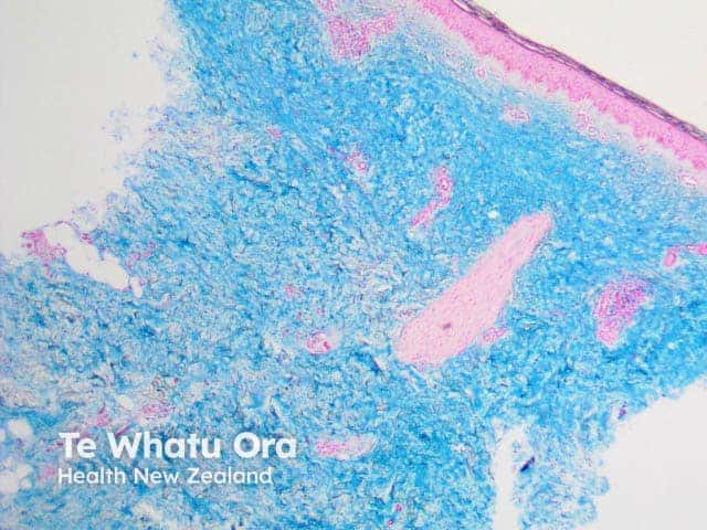
Alcian blue |
Blue; common mucin stain Image is pretibial myxoedema |
 |
Aldehyde fuchsin |
Purple/black; used to stain beta cells in the pancreas |
|
Alkaline phosphatase |
Red/blue; used for endothelial tissue |
|
Bielshowsky stain |
Black; used for neural plaques and tangles |
|
Congo red |
Red; typical for staining amyloid fibres Image is cutaneous amyloid |
 |
Crystal violet |
Violet; can stain glia and neurons |
|
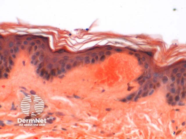
Eosin |
Pink/orange/red; typical for general staining when combined with haematoxylin Image is of normal skin |
 |
Fontana-Masson |
Black/pink or red; stains melanin |
|
Giemsa |
Blue/violet/pink; commonly used in blood or bone marrow smears |
|
Haematoxylin |
Blue/purple; standard for general staining when combined with eosin Image is of normal skin |
|
Luna stain |
Purple/black; can stain mast cells and elastin |
|
Nissl |
Blue; stains the rough endoplasmic reticulum in neurons |
|
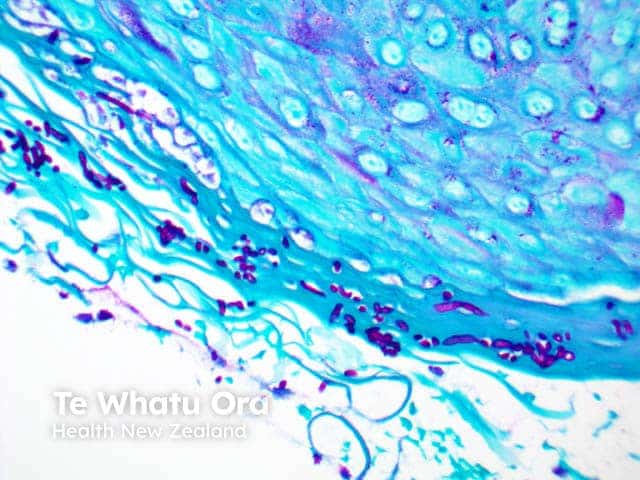
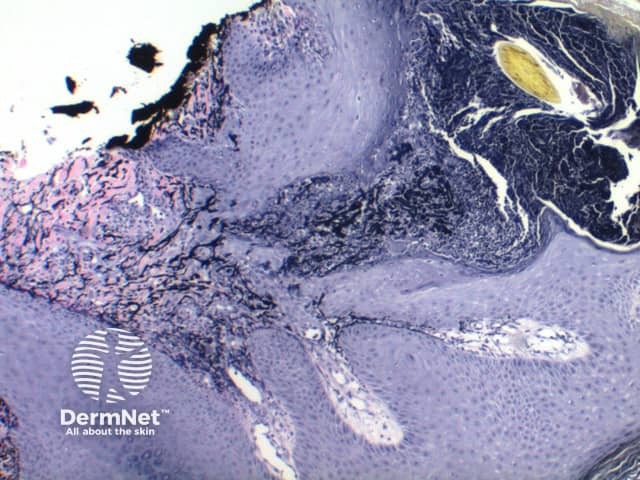
Period Acid Schiff (PAS) |
Red/magenta; used to stain glycogen, basement membranes, reticular fibres, cartilage, glycoproteins, glycolipids and mucins in tissues. Image shows superficial fungal elements due to candida infection. |
 |
Red Oil 3 |
Red; used to stain fat emboli |
|
Reticulin stain |
Blue/black; stains reticular fibres |
|
Sudan black |
Brown-black; stains myelin tissue |
|
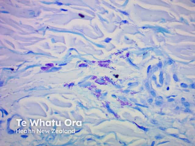
Toluidine blue |
Blue; stains mast cell granules Image is of urticaria pigmentosa |
 |
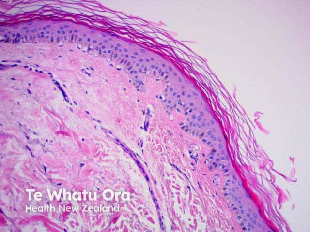
van Gieson |
Red/blue/yellow; used to study blood vessels and skin, can stain collagen, nucleus, red blood cells, cytoplasm Image is of elastosis perforans serpiginosa |
 |
What are the advantages and disadvantages of conventional histology?
The advantages of histology and histological staining are:
- It is one of the least expensive morphological methods
- Allows for the study of large sections of the tissue sample
- Can be collected relatively quickly and with little or no risk to the patient
- Gives clinicians an essential diagnostic tool and allows them the ability to examine the internal architecture of various cells and tissues.
The disadvantages of histology and histological staining include:
- Preparation of the slides using the paraffin technique can be time-consuming; frozen slides are faster to prepare, but this can affect the resolution, especially when using light microscopy
- The method is subject to human error during the preparation and analysis of slides
- This type of staining can be less specific than speciality staining techniques like immunohistochemical (IHC) staining.
- It can sometimes be difficult to identify specific types of cells with this technique, which can affect its value as a diagnostic tool.
References
- Cellular imaging, Southwest Environmental Health Sciences Center, University of Arizona, 2015.
- Histology stains. Histology World. 2014.
- Peckham, A. Histology guide. Faculty of Biological Sciences, University of Leeds, 2003.
- Routine and special staining. Leica Biosystems. 2014.
On DermNet