Introduction Histology Special studies Differential diagnoses
Keratoelastoidosis marginalis presents as symmetric, hyperkeratotic, linear plaques along the lateral aspects of the hands. It has otherwise been reported as digital papular calcific elastosis of the hands, and degenerative collagenous plaques of the hands.
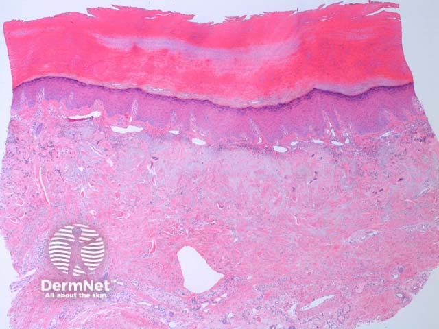
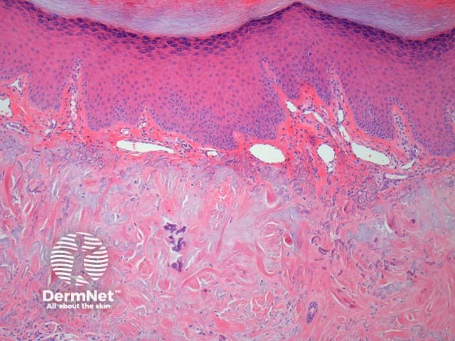
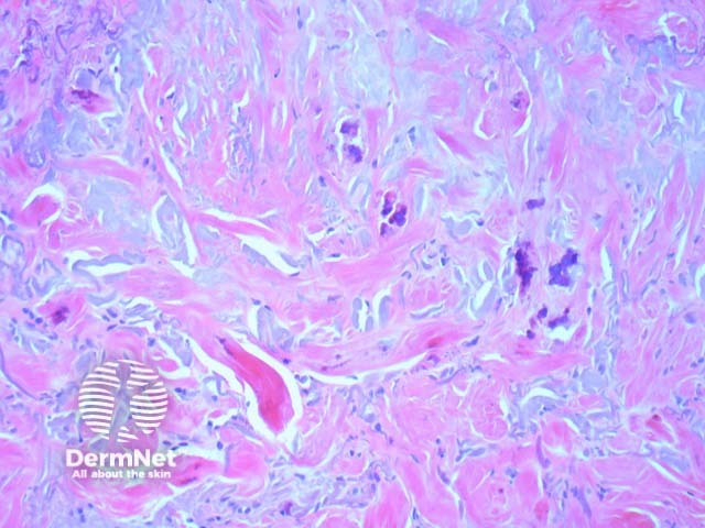
Histologic findings in keratoelastoidosis marginalis include epidermal acanthosis with some loss of the rete pattern associated with overlying hyperkeratosis (figure 1). There are thick acellular collagen bundles in the dermis (figure 2). In addition, there are fragmented degenerative elastic which may form elastotic masses. These masses sometimes calcify (figure 3).
Elastic stains will show a reduction of elastic fibres. The residual elastic fibres are disorganised or can form masses
Dermal elastosis – the dense fibroplasia and clinical features help to distinguish this disorder from regular dermal elastosis.

