

Lichenoid drug eruption pathology — extra information
Introduction Histology Special studies Differential diagnoses
Introduction
Lichenoid drug reactions are induced by a medication or another exogenous source which can mimic other lichenoid dermatoses clinically and histologically.
Histology of lichenoid drug eruption
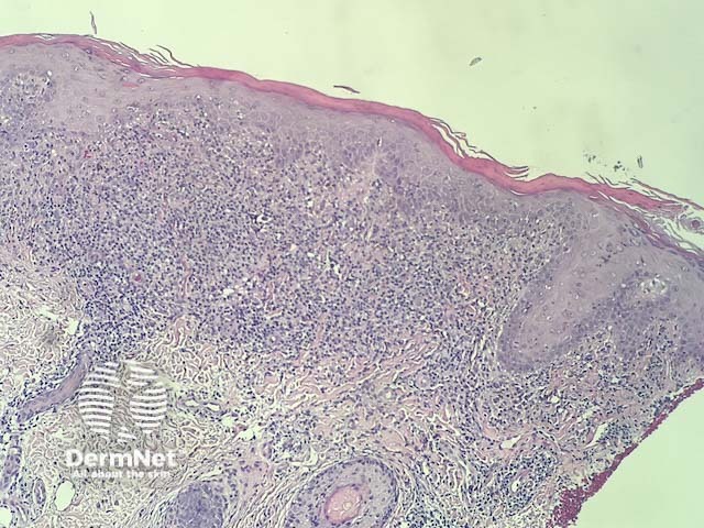
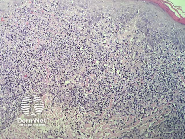
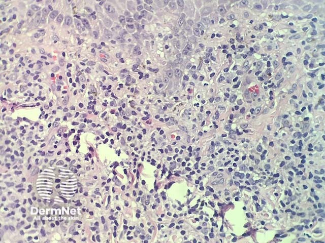
In lichenoid drug reactions the pathology is nearly identical to lichen planus. There is a dense, band-like lymphocytic infiltrate in dermis that obscures dermoepidermal junction, cytoplasmic vacuolisation of basal keratinocytes and apoptotic keratinocytes that degenerate into colloid bodies (figures 1,2). In addition, there are usually eosinophils in the infiltrate (best seen in figure 3).
Special studies for lichenoid drug eruption
None are generally needed.
Differential diagnosis for lichenoid drug eruption
Other diagnoses to be considered include any lichenoid dermatosis. The key differential is lichen planus. Clinical correlation can be very useful. Key histologic features seen in lichenoid drug eruptions, that are not common in idiopathic lichen planus, include the presence of eosinophils and the presence of prominent parakeratosis.
References
- Lichenoid drug eruption. Clues to diagnosis in dermatopathology. Derm101.com (accessed 10 September 2018).
On DermNet
Other websites
- Lichenoid dermatitis — PathologyOutlines.com
Books about skin diseases