

Lymphangioma circumscriptum pathology — extra information
Introduction
Lymphangioma circumscriptum presents on the skin surface as grapelike groups of thin-walled, translucent, lymph-filled vesicles, often compared with frog spawn. Haemorrhage within the lesions can create a deep red or black appearance.
Histology of lymphangioma circumscriptum
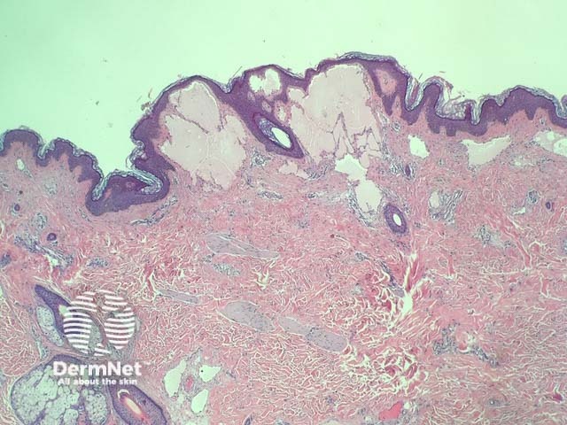
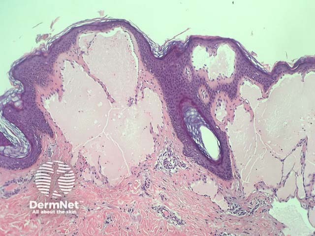
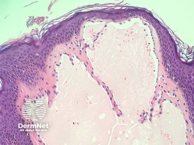
In lymphangioma circumscriptum, histopathological examination reveals acanthosis and hyperkeratosis of epidermis (figure 1). Within the papillary and reticular dermis, there are dilated lymphatic channels containing eosinophilic proteinaceous material in the papillary dermis (figures 2,3).
None are usually needed. Lymphatic architecture can be highlighted with immunohistochemical markers such as D2-40.
Differential diagnosis for lymphangioma circumscriptum
Angiokeratoma can look very similar to lymphangioma circumscriptum but are composed of blood vessels containing blood rather than lymphatics containing lymphatic fluid.
References
- Sinha A, Phukan JP, Jalan S, Pal S. Lymphangioma circumscriptum of the vulva: Report of a rare case. Journal of Mid-Life Health. 2015;6(2):91–3. doi:10.4103/0976-7800.158968. PubMed Central
On DermNet