Are you a healthcare professional
GO TO DERMNET PRO
Main menu
Common skin conditions
NEWS
Join DermNet PRO
Read more
Quick links
Skin checker
Try our skin symptom checker
Topics A-Z
Mohs surgery images
Treatments
Author: Tom Middelburg, MD PhD, dermatologist and Mohs surgeon, Christchurch, New Zealand, May 2017.
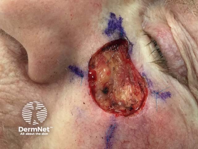
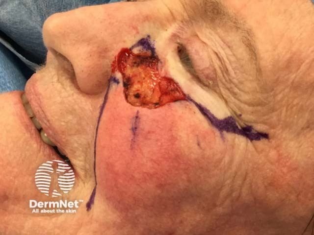
Example of a Mohs procedure
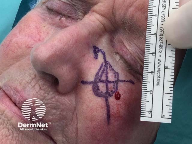
This infiltrative basal cell carcinoma required 5 Mohs stages.
Clinically visible basal cell carcinoma plus margin is outlined, a grid is created as part of the ma
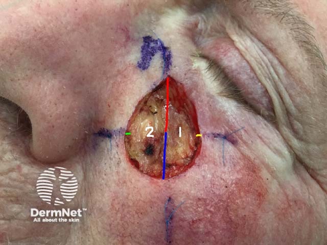
Tissue sample is excised and cut into two specimens which are colour code
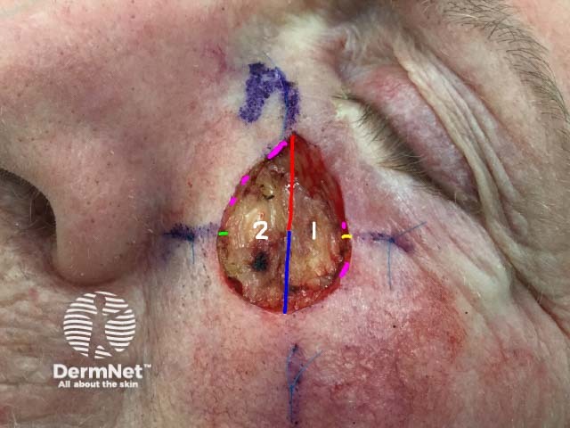
After microscopic examination of histological slides any areas of residual tumor are recognised and
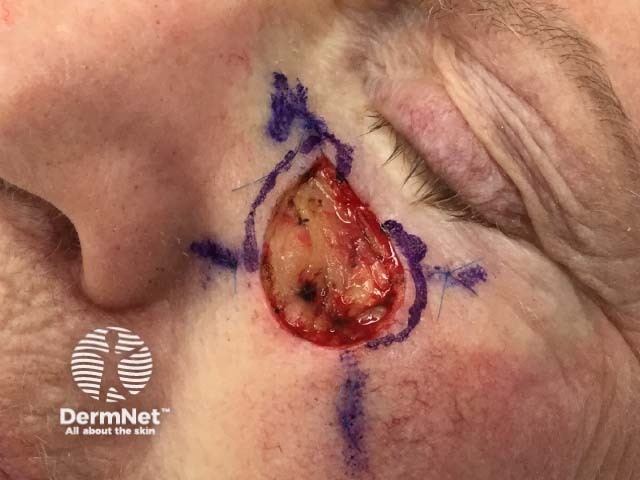
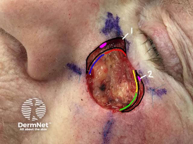
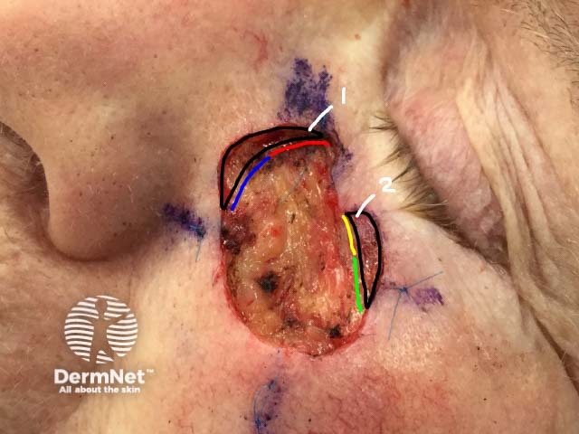
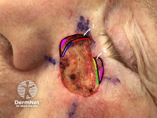
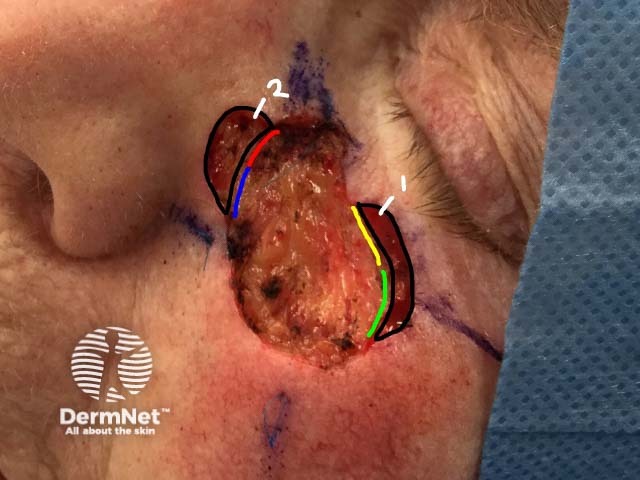
In the second Mohs stage the area to be excised is marked on the patient
The marked areas are excised
The two tissue samples are colour coded
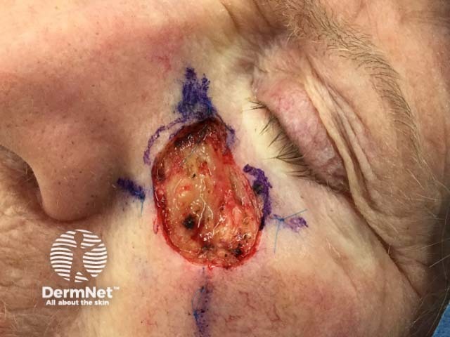
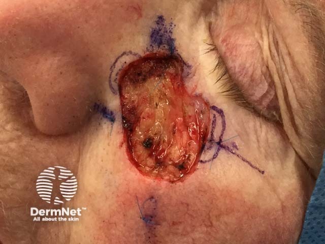
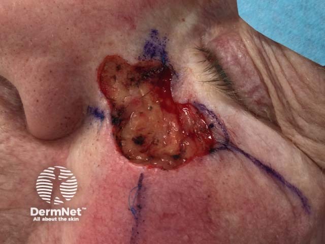
In the third Mohs stage the area to be excised is marked on the patient
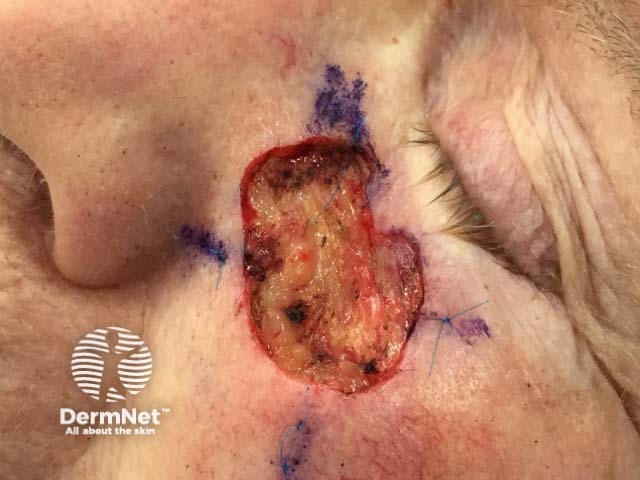
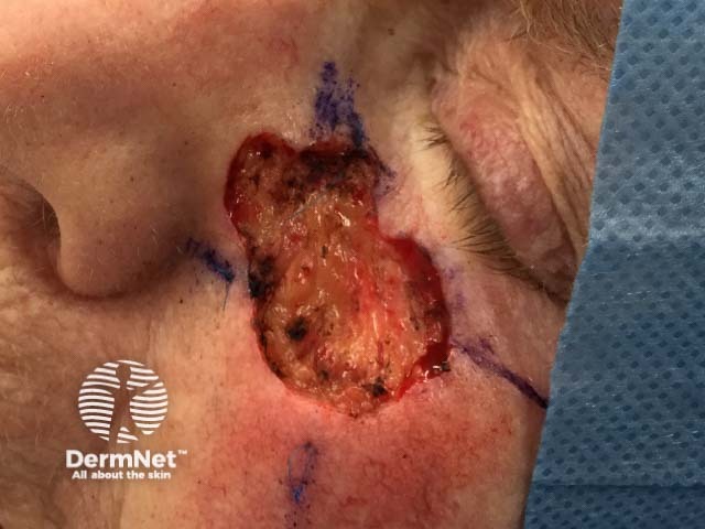
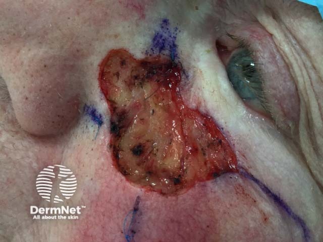
In the fourth Mohs stage the area to be excised is marked on the patient
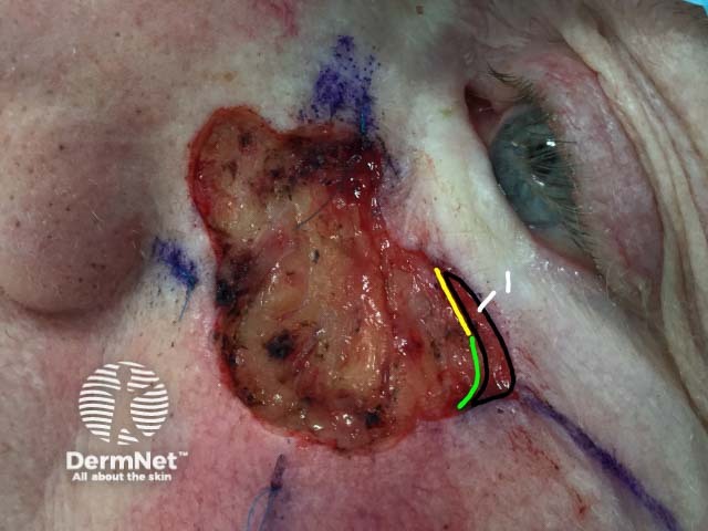
In the fifth Mohs stage the area to be excised is marked on the patient
The marked area is excised
The area is free of tumour cells
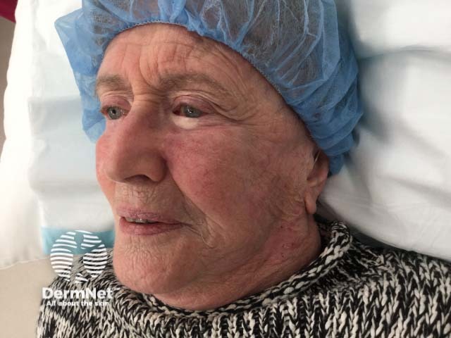
The wound is reconstructed, in this case by a cheek advancement flap
Two months after the Mohs procedure