Introduction Histology Special studies Differential diagnoses
Neurofollicular hamartoma presents as a smooth flesh-coloured papule on the face.
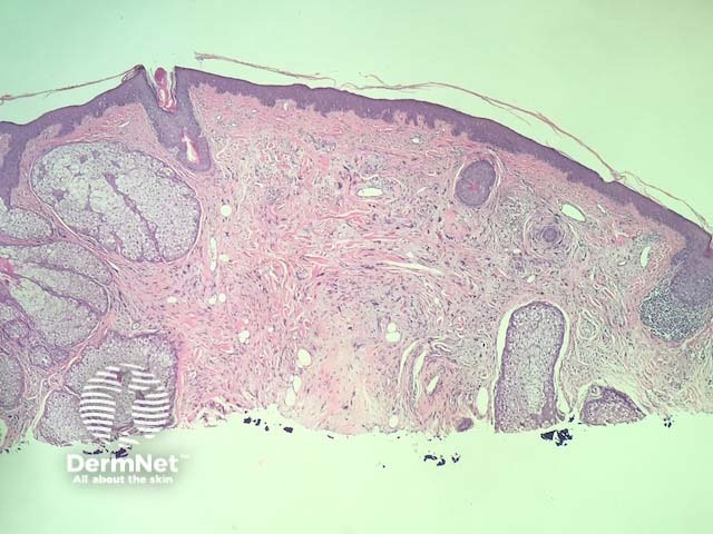
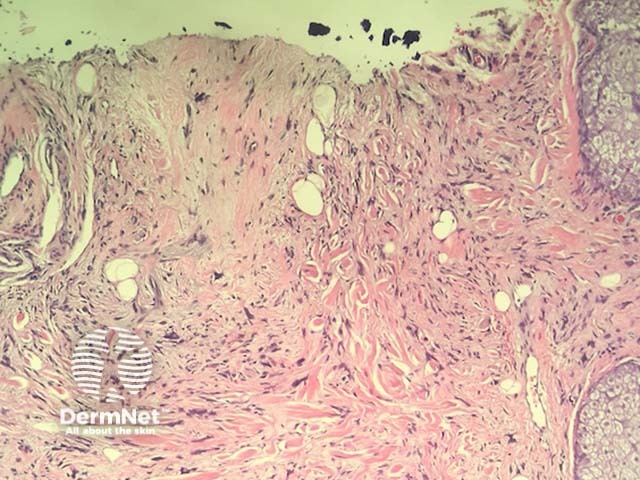
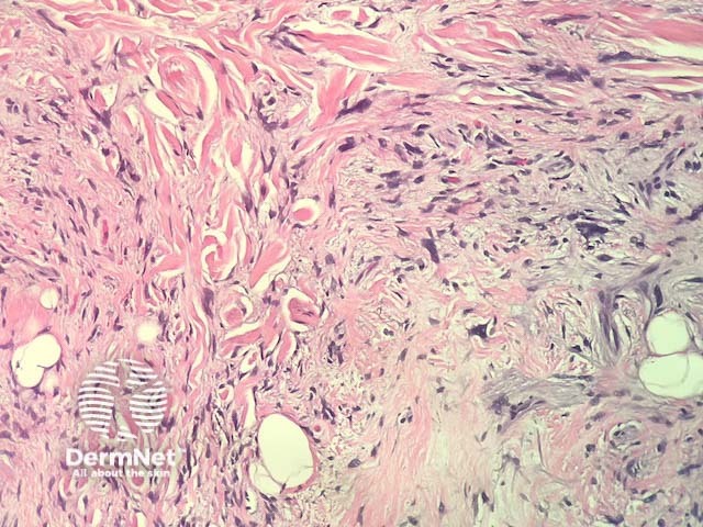
In neurofollicular hamartoma, the histopathology shows a nodular dermal spindle cell proliferation surrounded by prominent sebaceous glands. The spindle cells are plump or wavy. The sebaceous proliferation surrounds the mesenchymal component (figures 1–3).
The spindle cells are positive for S100, CD34 and Factor XIII.
Other diagnoses to be considered include:
Requena L, Yus ES, Simón P, del Rio E. Induction of cutaneous hyperplasias by
altered stroma. Am J Dermatopathol. 1996 Jun;18(3):248–68. PubMed PMID: 8806959.

