

Nipple papillomatosis — extra information
Introduction Demographics Clinical features Complications Diagnosis Differential diagnoses Treatment
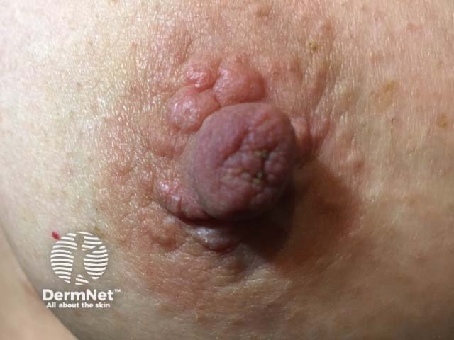
What is nipple papillomatosis?
Nipple papillomatosis is characterised by the benign growth of numerous papillomas arising from the lactiferous duct epithelium of the nipple. It is also known as florid papillomatosis of the nipple, nipple adenoma, and erosive adenomatosis.
Who gets nipple papillomatosis?
Nipple papillomatosis is rare. It mainly occurs in women aged 35–55 years old, but it can occur at any age. It occasionally affects men.
What are the clinical features of nipple papillomatosis and how is it diagnosed?
Nipple papillomatosis presents as a palpable mass or swelling on the surface of the nipple. It may be discovered incidentally during a breast examination.
Other clinical features include:
- Erosions
- Discharge of serous or bloody fluid
- Itching or tenderness.
What are the complications of nipple papillomatosis?
Nipple papillomatosis is harmless. As for any fibrocystic change within the breast tissue, there are reported associations with a slightly increased risk of breast cancer when compared with the general population, although this risk is still very low.
Syringomatous adenoma is a variant of nipple papillomatosis, distinguished on histology by the absence of intraductal epithelial hyperplasia and oval, elongated ducts. Syringomatous adenoma tends to be infiltrative and locally invasive.
How is nipple papillomatosis diagnosed?
A skin biopsy may be performed to confirm the diagnosis of nipple papillomatosis.
Histologically, nipple papillomatosis shows proliferating ductal structures extending into the breast stroma, lined by a double layer of epithelium. Other histological features include the presence of keratin-filled cysts and tiny apical cell snouts.
Breast X-ray, mammography or ultrasound examination may be performed to exclude an underlying tumour.
What is the differential diagnosis for nipple papillomatosis?
The differential diagnosis for nipple papillomatosis includes:
- Tubular and lactating adenomas (benign growths of epithelial cells of the mammary glands)
- Nipple eczema, often due to atopic eczema — which may result in oozing, crusting and scaling
- Paget disease of the breast
- Hyperkeratosis of the nipple and areola.
What is the treatment for nipple papillomatosis?
Where practical, nipple papillomatosis is treated by complete excision of the lesion. This is a minor procedure usually performed under local anaesthetic. If the entire affected area is not removed, the affected area should be reviewed from time to time, especially if the appearance changes.
References
- Champion RH, Burton JL, Burns DA, Breathnach SM (eds). Rook / Wilkinson / Ebling: Textbook of dermatology, 6th edn. Volume 4. Oxford: Blackwell Science; 1998.
- Salemis NS. Florid papillomatosis of the nipple: a rare presentation and review of the literature. Breast Dis. 2015; 35(2): 153-6. DOI: 10.3233/BD-150397. PubMed
- Halls S. Benign atypical ductal hyperplasia, intraductal papilloma. June 2017. Available at www.breast-cancer.ca/3b-hyperplasia/ (accessed 17 June 2017).
- Hall S. Nipple adenoma. June 2017. Available at www.breast-cancer.ca/nip-ade/ (accessed 17 June 2017).
- Hall S. Papilloma and papillomatosis. June 2017. Available at www.breast-cancer.ca/papily/ (accessed 16 June 2017).
On DermNet
- Erosive papillomatosis of the nipple pathology
- Mammary Paget disease
- Nipple eczema
- Scaly skin conditions
- Hyperkeratosis of the nipple and areola
- Nipple papillomatosis
- Nipple skin problems
Other websites
-
Nipple adenoma / florid papillomatosis of nipple — Pathology Outlines, July 2014 (accessed 17 June 2017).