

Pityriasis versicolor pathology — extra information
Histology of pityriasis versicolor
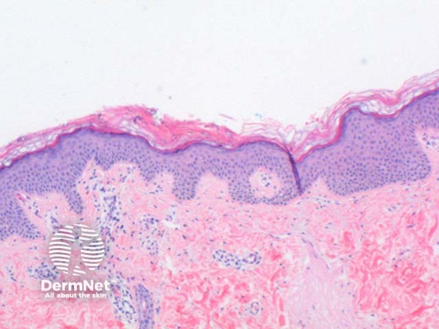
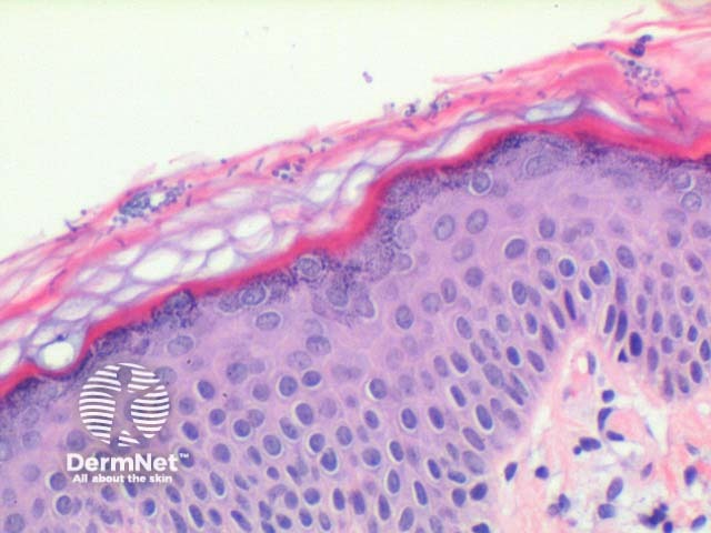
The low power view of pityriasis versicolor is suggestive of a mild superficial perivascular inflammation pattern (Figure 1). The epidermis may show mild hyperkeratosis and acanthosis (Figure 2). Elongation of the rete ridges and mild increased basal layer pigmentation as seen in acanthosis nigricans can sometimes be observed.
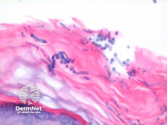
In pityriasis versicolor, the organisms are seen almost exclusively within the stratum corneum, and are readily seen on haematoxylin and eosin sections (Figures 2, 3). The characteristic spores and short ‘cigar-butt’ hyphae of Malassezia furfur are likened to spaghetti and meatballs (Figure 4). A mild superficial perivascular lymphocytic infiltrate, rarely with plasma cells, is seen.

Special stains in pityriasis versicolor
Periodic acid-Schiff (PAS) and methenamine silver stains can improve the visualisation of the organisms within the stratum corneum.
Differential diagnosis of pityriasis versicolor
While the presence of the characteristic fungal forms in the appropriate clinical setting is diagnostic, when there are few fungal elements consideration should be given to:
- Confluent and reticulated papillomatosis of Gougerot and Carteaud: While similar epidermal changes can be seen, the degree of hyperkeratosis and acanthotic downgrowth is usually more pronounced. Typically, few or no fungal elements are seen.
- Acanthosis nigricans: Occasional incidental (non-pathogenic) spore forms can be seen in the stratum corneum.
References
- Skin Pathology (2nd edition, 2002). Weedon D
- Pathology of the Skin (3rd edition, 2005). McKee PH, J. Calonje JE, Granter SR
On DermNet