

Proliferating epidermoid cyst pathology — extra information
Introduction Histology Special studies Differential diagnoses
Introduction
Proliferating epidermoid cyst has been poorly defined in the literature. The regular epidermoid cyst should be seen in at least part of the lesion in addition to an epidermal proliferation.
Histology of proliferating epidermoid cyst
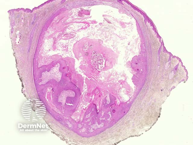
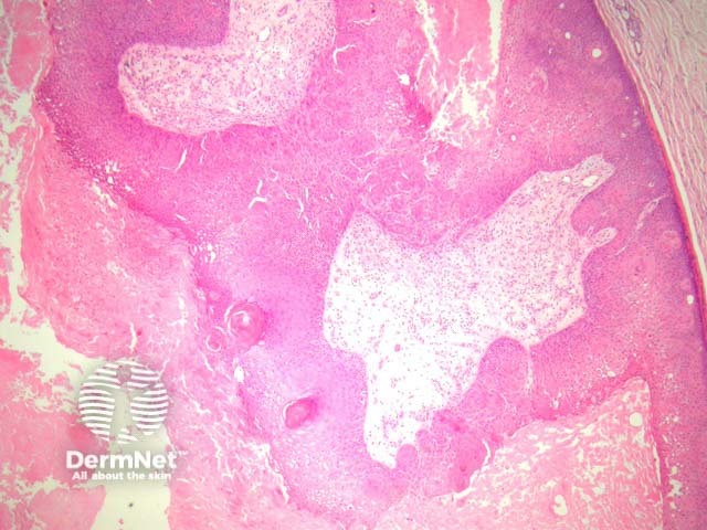
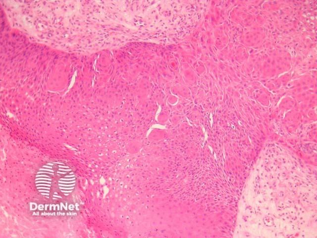
Sections show a cyst in the dermis with a proliferating epidermal component (figures 1, 2). Characteristically, the proliferative areas are made up of bland squamous epithelium with striking squamous eddies (figures 2, 3, 4). These eddies are whorles of maturing squamous epithelium and are exactly the same as those seen in irritated seborrheic keratoses or inverted follicular keratoses.

Special studies for proliferating epidermoid cyst
None are needed.
Differential diagnosis of proliferating epidermoid cyst pathology
HPV-related epidermal cysts – These have a hyperplastic lining with viropathic nuclear and cytoplasmic changes
Cystic squamous cell carcinoma – Must be considered if there are nuclear atypia and adjacent infiltration into the surrounding dermis. This can be a challenging differential when cysts have partially ruptured or there is extensive proliferation.
References
- Pathology of the Skin (Fourth edition, 2012). McKee PH, J. Calonje JE, Granter SR
On DermNet