

Treated melanoma pathology — extra information
Introduction
Histology
Special studies
Differential diagnoses
Introduction
New therapies targeting BRAF and MEK have emerged as the key component for the treatment of BRAF-mutant metastatic melanoma.
Histology of treated melanoma
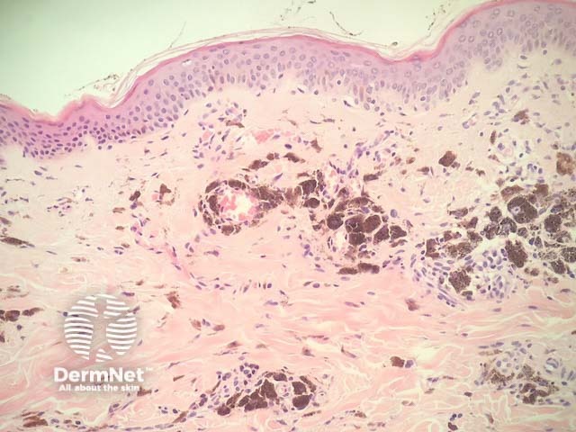
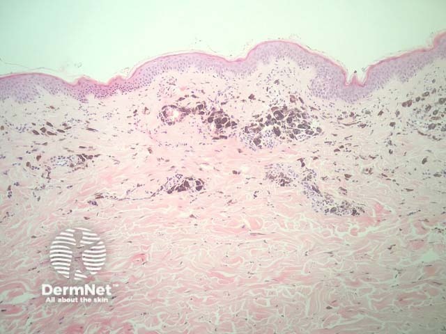
In melanoma treated with targeted BRAF and MEK therapy, the histopathology usually shows evidence of extensive regression with numerous melanophages and inflammatory cells within areas which previously housed melanoma (figures 1,2).
Special studies for treated melanoma
Given the extensive melanin deposition and inflammation, it can be difficult to appreciate viable residual melanoma. Bleaching the section can help reduce this difficulty. Immunohistochemistry with Sox-10 can help identify residual melanoma.
Differential diagnosis for treated melanoma
It is important to have the correct clinical context and a history of targeted therapy. Without this history, the histopathology could precisely resemble natural regression in melanoma or regression of other tumours such as pigmented basal cell carcinoma or other pigmented lesions.
References
- Iams WT, Sosman JA, Chandra S. Cancer J 2017; 23: 54–8. DOI: 10.1097/PPO.0000000000000242. PubMed
On DermNet
- Targeted cancer therapies
- Melanoma
- Metastatic melanoma
- Melanoma pathology
- Skin lesions, tumours and cancers
- Dermatopathology glossary
- Dermatopathology index