Author(s): Dr Zsófia Sára Hermann, Senior House Surgeon, Health New Zealand Capital, Coast and Hutt Valley, New Zealand; Hon. Prof. Amanda Oakley, Dermatologist, Health New Zealand Waikato (2025)
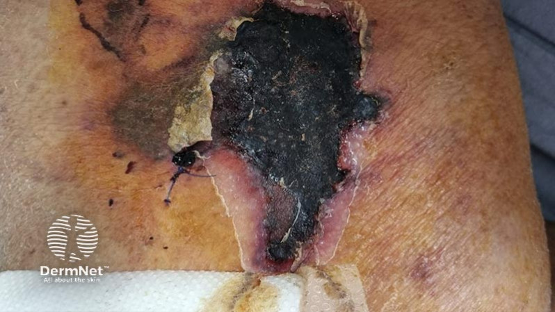
A 65-year-old man presents to the emergency department with a painful, necrotic wound on both of his thighs which developed over the course of a few days. He experienced associated malaise, but no fevers.
He has a background of type 2 diabetes mellitus, and chronic renal failure requiring dialysis due to multiple myeloma.
There is a jagged area of skin necrosis with an overlying slough.
The diagnosis was calciphylaxis, which was confirmed by biopsy at the edge of the wound. It showed calcification within the lumen of the dermal small arteries. Calciphylaxis is probably better termed calcific uraemic arteriolopathy. It is characterised by calcification and thrombosis of skin arterioles, which results in tissue ischaemia, necrosis, and ulceration of the skin and subcutaneous tissues (occlusive vasculopathy). Biopsies of wounds secondary to calciphylaxis are often inconclusive due to sampling error, therefore many cases are diagnosed clinically.
The cause of calciphylaxis is not entirely understood.
Calciphylaxis is most common in patients who have chronic renal disease requiring dialysis.
It can also occur in people with normal renal function (termed normo-renal calciphylaxis), and risk factors include diabetes mellitus, female sex, Caucasian race, obesity, sudden weight loss and the use of warfarin.
It may also occur in those predisposed to higher serum calcium levels (such as malignancy and primary hyperparathyroidism).
Calciphylaxis in association with chronic renal disease has a mortality of 60–80%. Sepsis is the leading cause of death due to secondary bacterial infection of the wounds. Patients with calciphylaxis also suffer from severe pain that may not respond to strong analgesics.
It is difficult to treat - in those being dialysed, altering the dialysate may be of benefit. Warfarin can be replaced with either heparin or a NOAC. Intravenous sodium thiosulphate may help solubilise the intra-arterial calcium.

