Author(s): Dr Stanley Leong, Dermatology and Paediatric Registrar, New Zealand (2025)
Reviewing dermatologist: Dr Ian Coulson
Edited by the DermNet content department
This 80-year-old woman was referred with a non-healing wound on her left lateral chest.
Four years ago, she had a chest drain inserted for a left-sided pleural effusion. Following removal of the chest drain, the wound never healed but slowly increased in size and started to bleed.
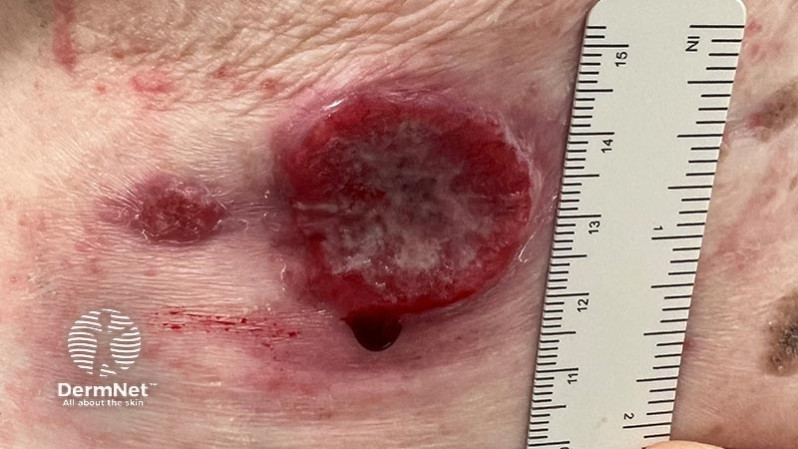
On examination, she was systemically well. She had 2 round lesions on her lateral chest, measuring 3cm and 1cm respectively. Both lesions were very friable and bled on contact. The borders of both lesions appeared slightly raised.
This is most likely a squamous cell carcinoma. Any non-healing wound should always be suspected as a cutaneous malignancy, and biopsied.
The histology result from this lady confirmed an undifferentiated squamous cell carcinoma. Tissue culture showed heavy growth of Staphylococcus aureus.
This case demonstrates the progression of a chronic, non-healing wound (originally from a chest drain) into a squamous cell carcinoma.
The treatment is usually surgical. Most cases are excised with a 3–10mm margin of normal tissue around a visible tumour. A flap or skin graft may be needed to repair the defect.
Other removal options include with poorer overall cure rates include:
Other treatment modalities include:

