Created 2008.
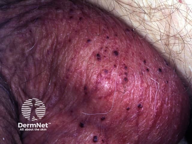
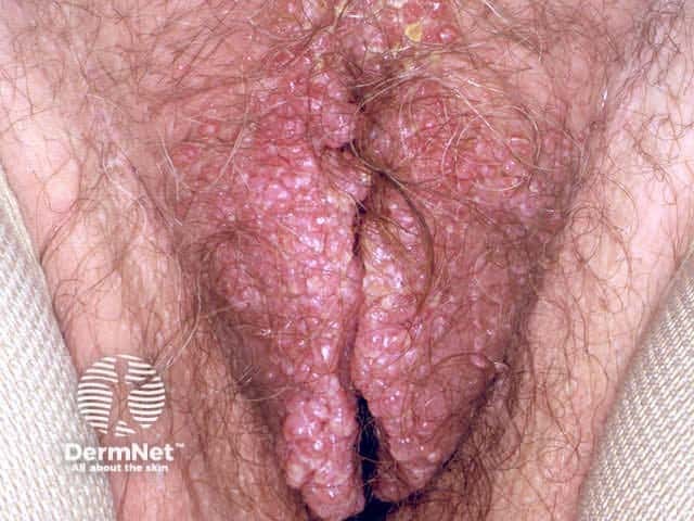
Angiokeratoma is a scaly vascular papule due to epidermal proliferation encircling dilated vessels. Angiokeratoma may be solitary or diffuse. Multiple lesions may be genital (Fordyce), acral (Mibelli) or rarely, generalised (Fabry disease: deficiency of ceramide trihexosidase resulting in deposition of glycosphingolipids).
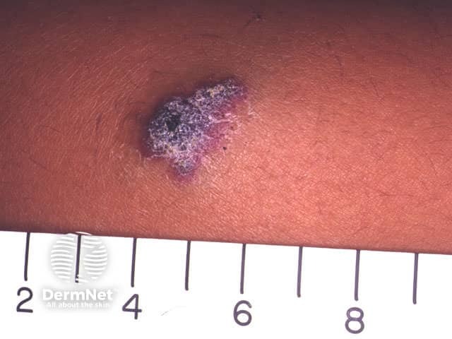
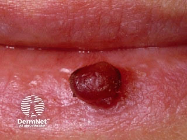
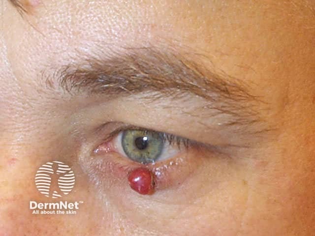
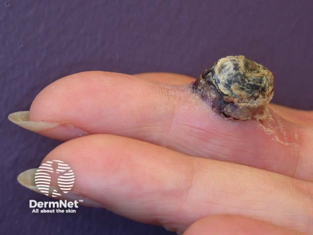
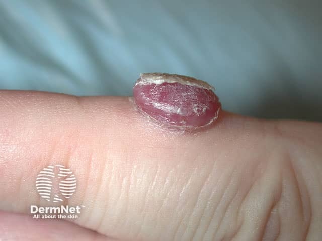
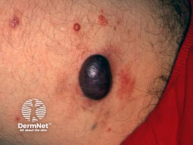
Pyogenic granuloma is one form of lobular capillary haemangioma. There is a characteristic collarette of skin around a juicy or friable red nodule that bleeds easily. Pyogenic granuloma sometimes follows minor trauma. Staphylococcus aureus is often isolated. Pyogenic granuloma is particularly common on lips and fingers, and during pregnancy and childhood.
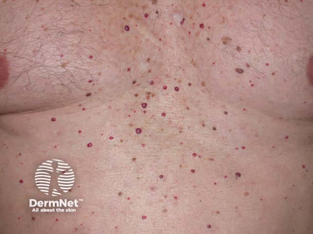
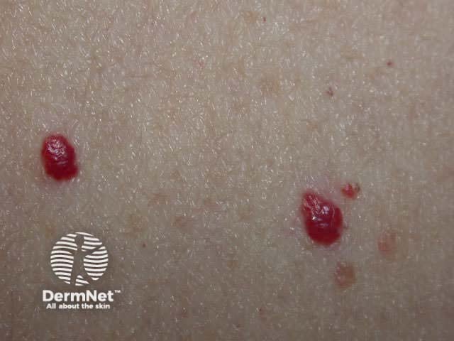
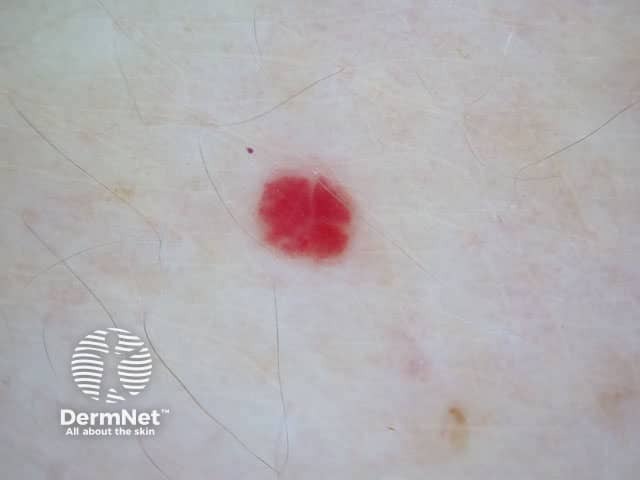
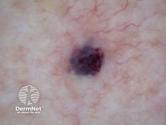
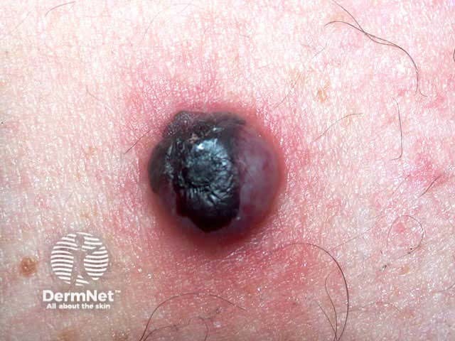
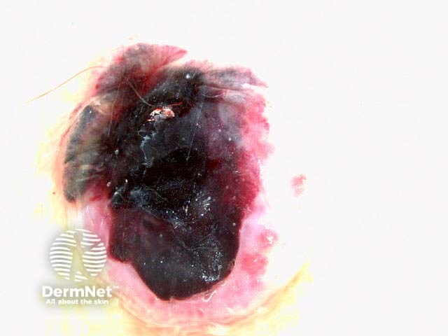
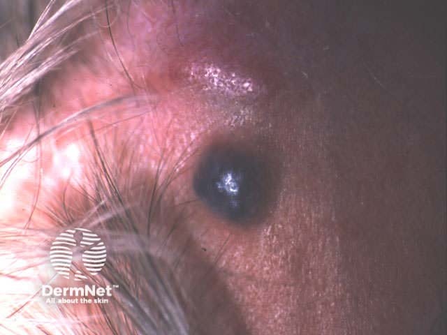
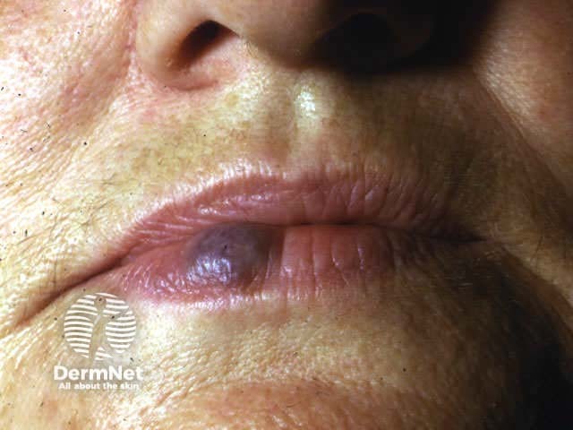
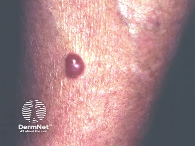
Cherry angiomas are extremely common benign red, blue, purple or almost black lesions occurring in middle age on the trunk. They can be easily distinguished from melanocytic lesions by dermoscopy, which shows red, blue or purple lacunes. Occasionally they become thrombosed and may fall off or persist as a firm bluish papule.
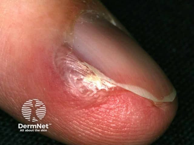
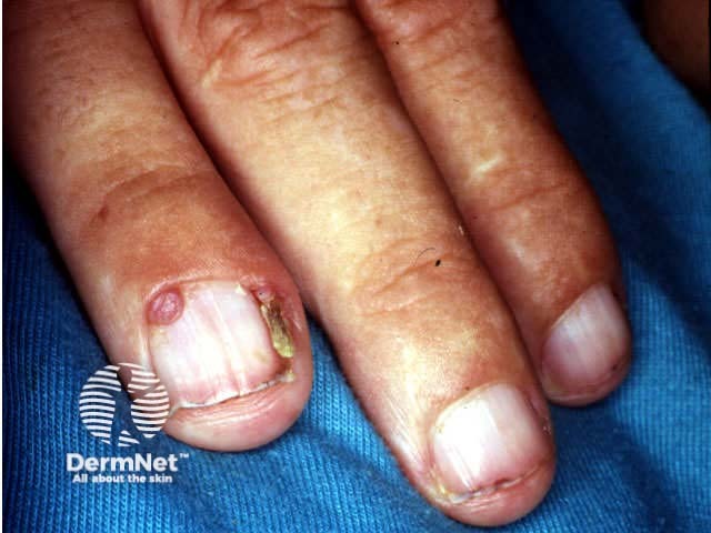
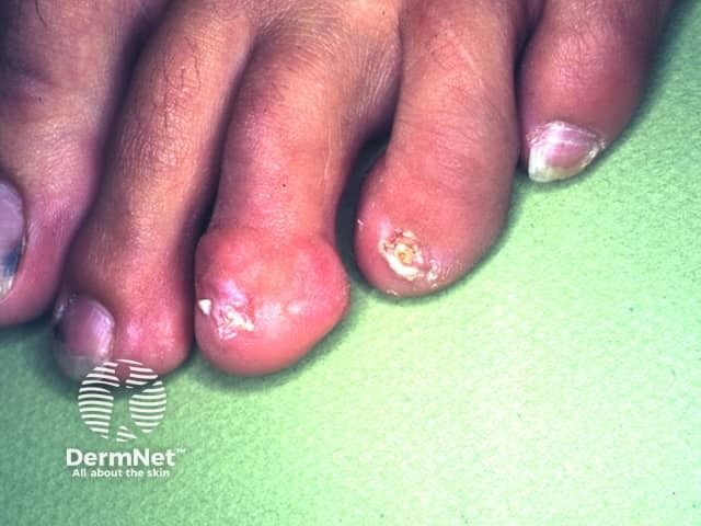
Glomus tumour usually presents as a painful subungual papule.
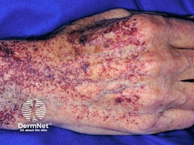
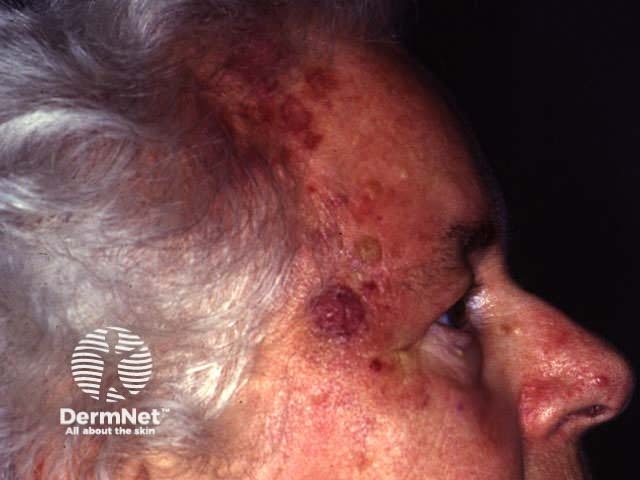
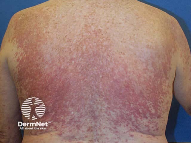
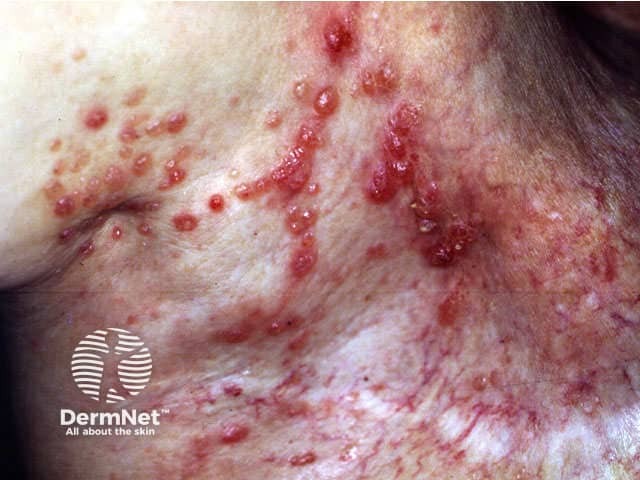
Angiosarcoma usually arises on the head and neck, or arise in areas of chronic lymphoedema such as after mastectomy. It presents as an advancing area of purpura and ecchymosis, usually in an elderly person. Prognosis is poor as the tumour is often multifocal so difficult to excise, and only partially sensitive to radiation.
Kaposi sarcoma (KS) is a low grade vascular malignancy related to human herpesvirus 8. There are four types.
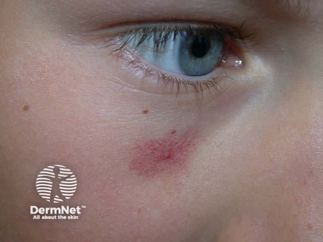
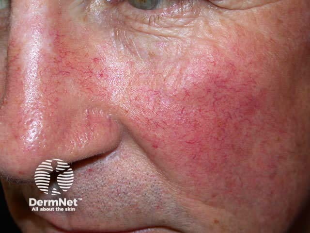
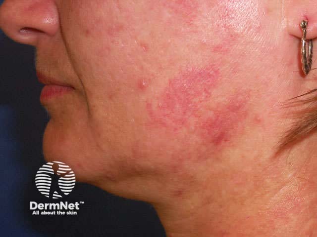
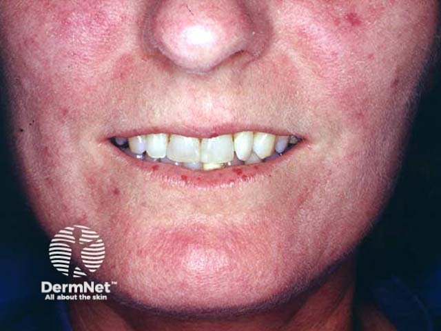
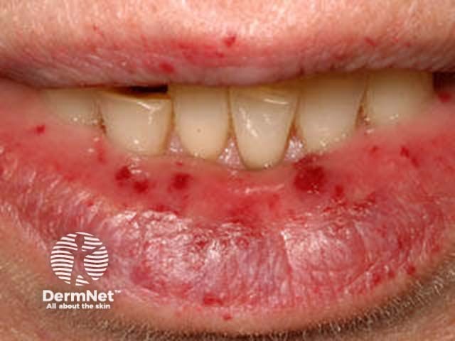
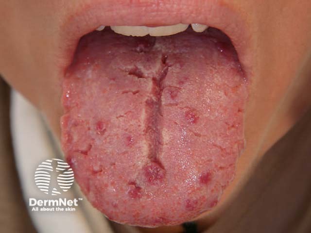
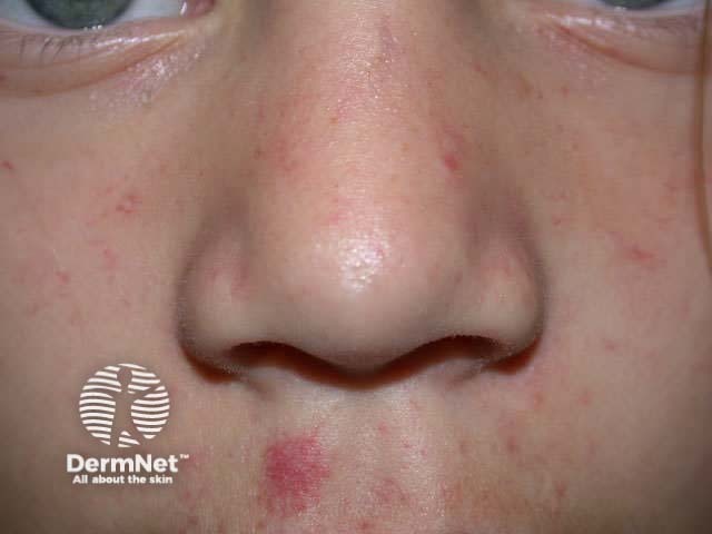
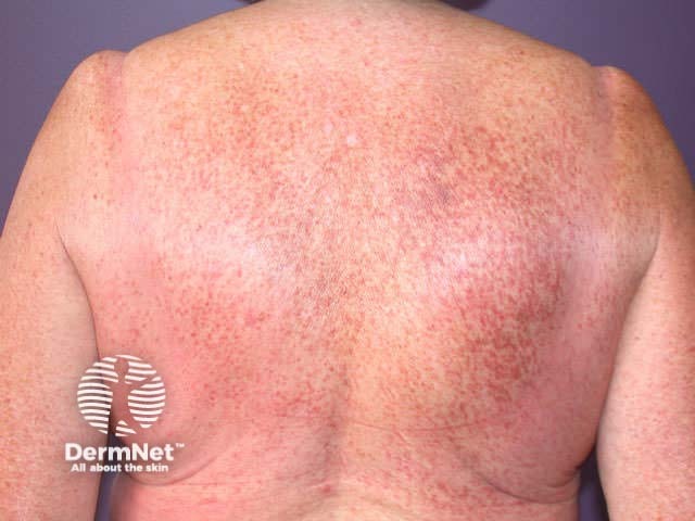
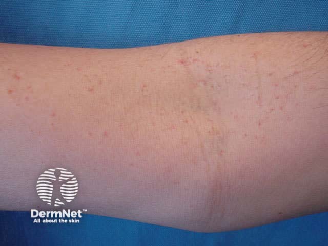
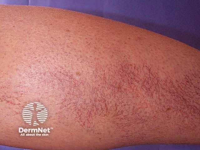
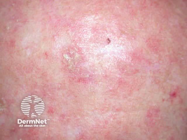
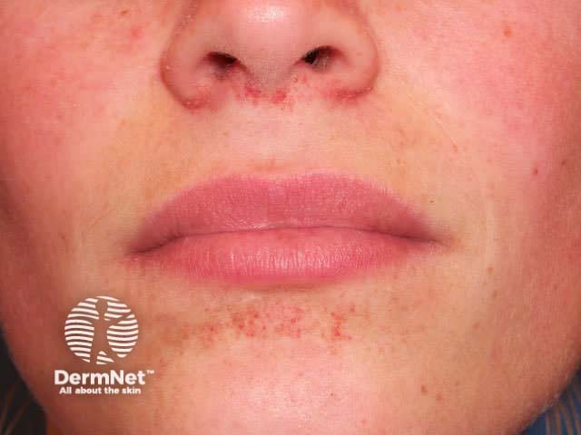
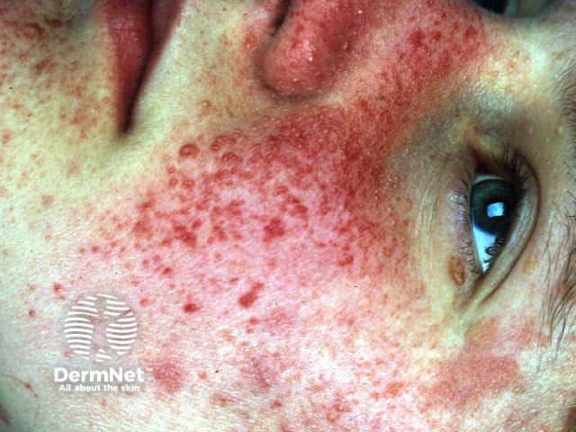
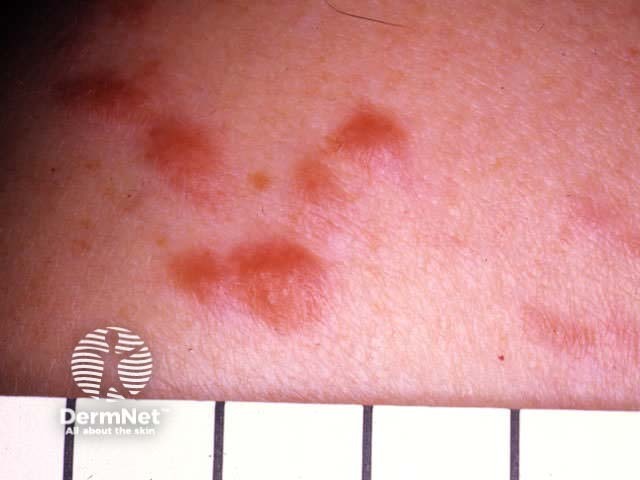
Acquired telangiectasia is common and affects vessels of various size. Some examples are illustrated.
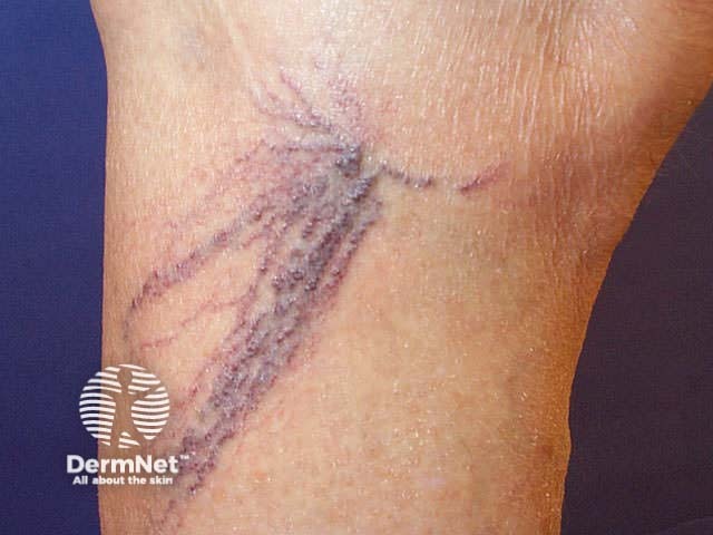
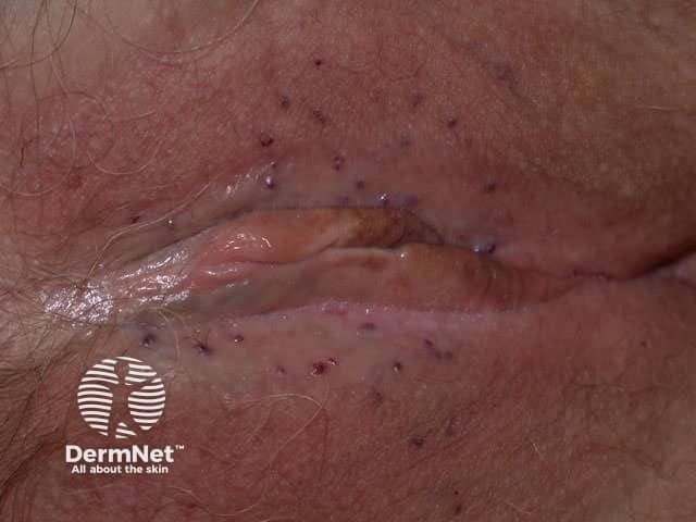
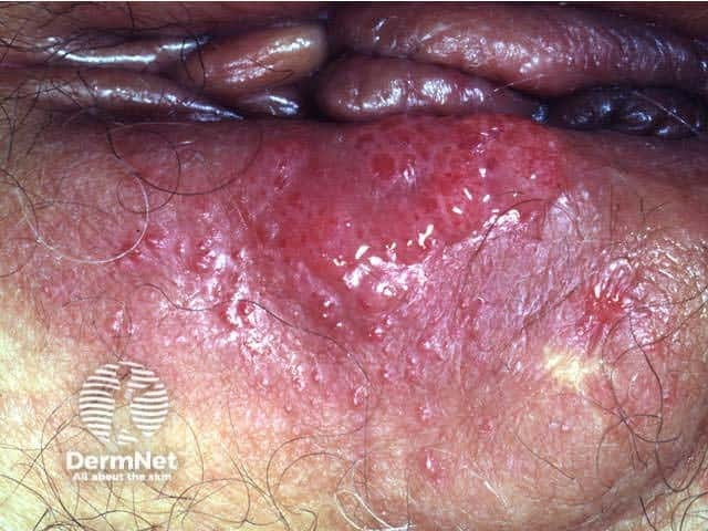
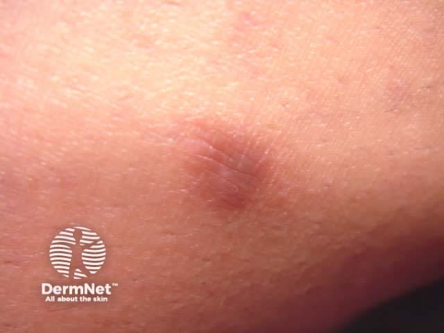
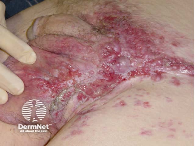
Acquired lymphangiectasia usually follows lymph node dissection, or traumatic injury interrupting lymphatic drainage, usually in the axilla or genital area. Frogspawn-like clear or haemorrhagic papules develop some time later and tend to cause distressing ooze.
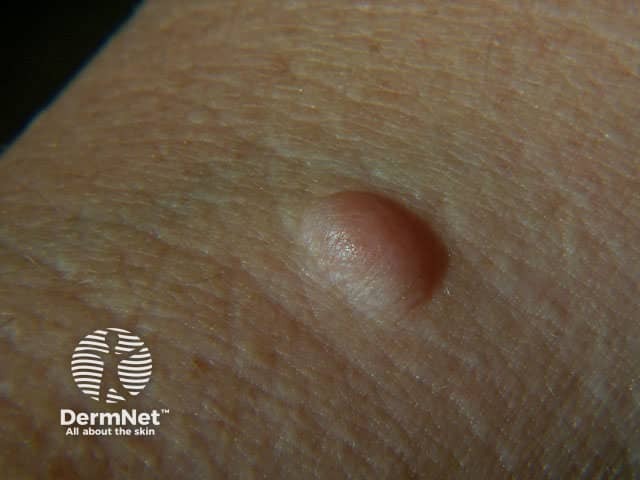
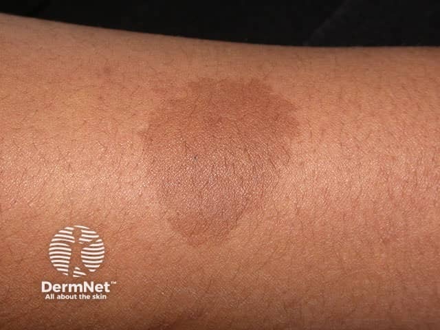
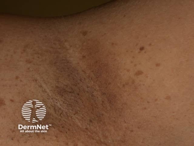
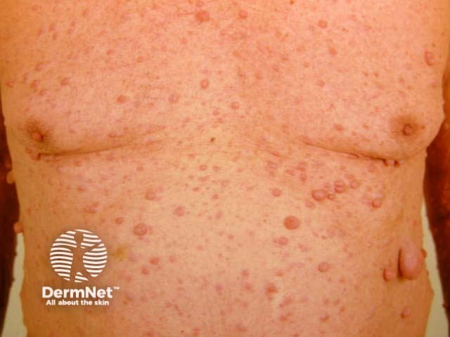
Neurofibromas are spindle cell tumours and present as soft to firm, single or multiple dermal nodules that may be pedunculated. Sometimes it is possible to invaginate the lesions because there is a defect in the dermis. There are diffuse, pigmented and plexiform variants. Plexiform neurofibroma is pathognomonic of NF1 neurofibromatosis; these may rarely undergo malignant transformation. Neurofibromatosis may also present with café au lait macules (>6), which are present at birth; axillary freckling; and other types of cutaneous neurofibroma, which arise later in childhood and adult life.
Other neural tumours include neurilemmoma, neuroma, granular cell tumour and neurofibrosarcoma (malignant schwannoma).
Merkel cell carcinoma (primary neuroendocrine carcinoma of the skin) is rare. It presents as a rapidly growing violaceous nodule that can ulcerate. It frequently recurs after excision; distant metastases develop in about 40% and 30% die of their disease within 5 years.
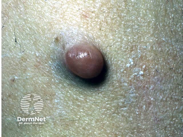
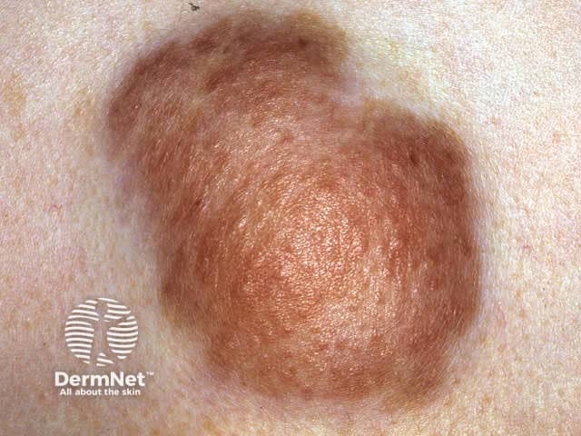
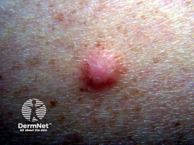
Dermatofibromas (fibrous histiocytomas) are common firm dermal papules or nodules caused by a proliferation of fibroblasts. They often follow insect bites, so most frequently arise on lower legs. They dimple on lateral compression (pinch or dimple sign).
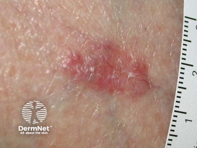
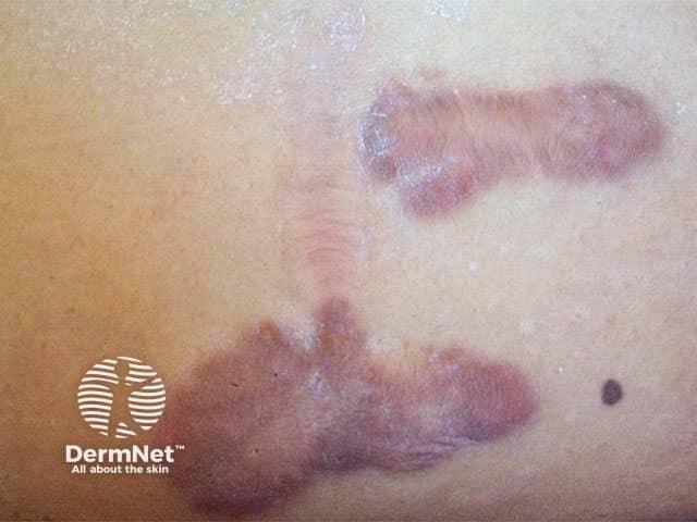
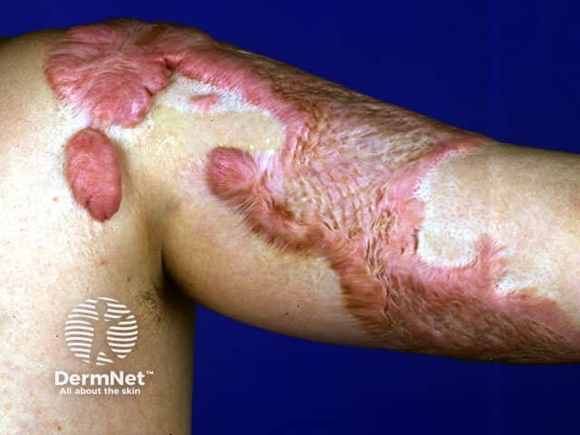
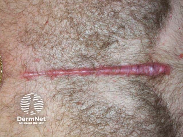
Keloids are scars with excessive bands of collagen.
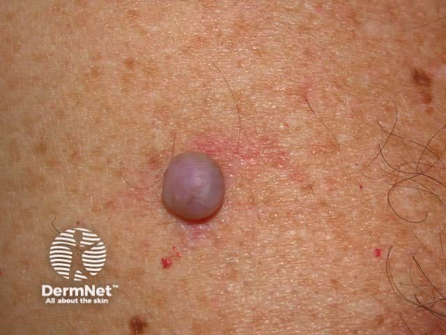
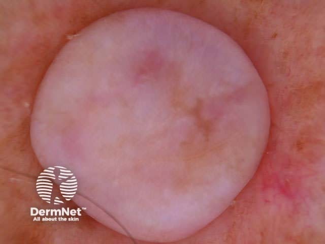
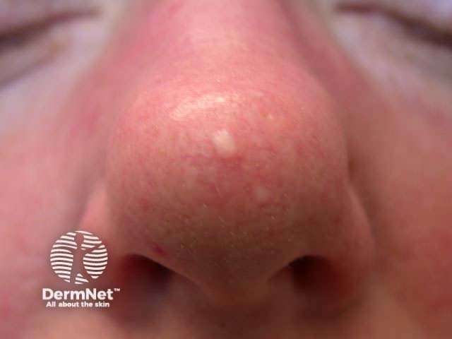
A solitary angiofibroma may also be called a fibrous papule or perifollicular fibroma. Fibrous papules are very common on the nose and although they look like intradermal naevi the lesion feels firm. They can be removed by shave excision or electrodessication. Multiple angiofibromas around the nose and cheeks are associated with tuberous sclerosis, appear during childhood and become increasingly prominent with age. These patients may also have periungual fibromas.
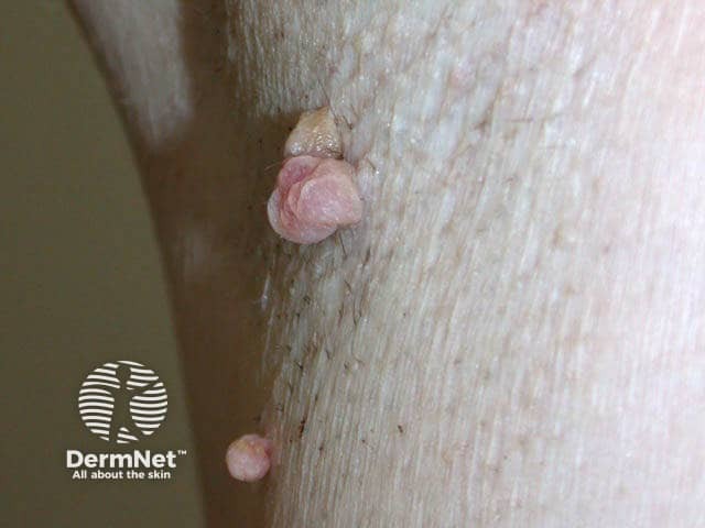
An acrochordon is a pedunculated skin tag filled with loose collagen.

Other benign and malignant fibrous lesions include:
Cutaneous lesions due to distant primaries are most often due to:
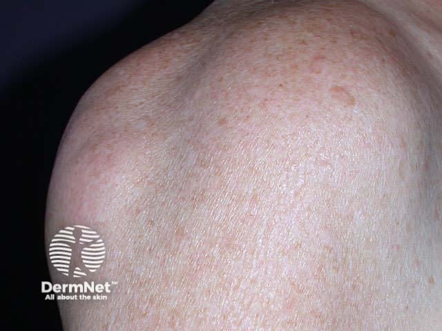
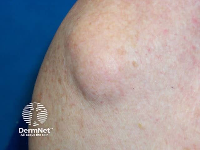
Lipomas are very common. They are an encapsulated proliferation of mature adipose tissue and present as solitary or multiple asymptomatic soft subcutaneous nodules. Multiple lesions are seen in association with various syndromes and may be familial.
Leiomyoma includes multiple piloleiomyomas, genital leiomyoma and angioleiomyoma. They usually present in young adults and may be painful or tender.
Uncommon skin tumours include:
Look for the skin lesions described in this section in the next twenty or so patients you see.
Information for patients
See the DermNet bookstore.

