DermNet provides Google Translate, a free machine translation service. Note that this may not provide an exact translation in all languages

Translate

Principles of dermatological practice

Structure of the epidermis

Created 2008.

Learning objectives

  • Name structural components of the normal epidermis and its appendages

Introduction

The skin of an adult occupies an area of 1.5 to 2 m2. It varies in thickness from 0.3 to several centimetres in thickness. The thinnest sites are the eyelids (a few cells thick) and scrotum. The thickest are the soles and palms (about 30 cells thick). The total weight of skin can reach 20 kg, about 16% of total body weight.

Skin is made up of:

  • Epidermis
  • Basement membrane zone
  • Dermis
  • Subcutaneous tissue.

Normal skin

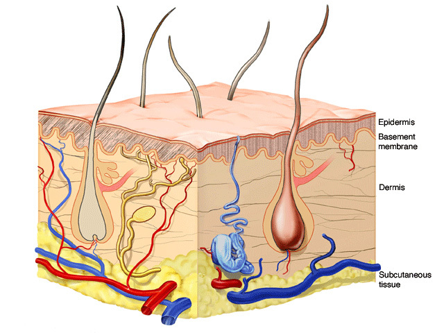
These layers are modified according to the needs of the specific area of the body. For example, the scalp is covered with thick hair, the palms have particularly thick epidermis and the face contains large numbers of sebaceous glands.

Each square centimetre of skin is said to have approximately:

  • 6 million cells
  • 5,000 sense end organs
  • 400 cm nerve fibres
  • 200 pain sensors
  • 100 cm blood vessels
  • 100 sweat glands
  • 15 sebum glands
  • 12 cold receptors
  • 5 hairs
  • 2 heat receptors

Acid mantle
Skin has an average pH value of 5.5, creating the acid mantle. This is the result of acidic substances such as amino acids, lactic acid and fatty acids in perspiration, sebum and the hormones. There are resident protective microflora (bacteria and yeasts) but the acid mantle repels pathogenic micro-organisms and reduces body odour.
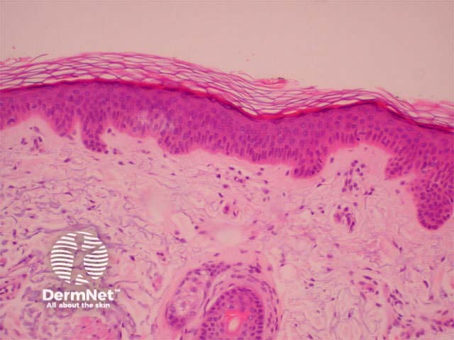
Epidermis
The epidermis is a dynamic structure acting as a semi-permeable barrier with a layer of flat anuclear cells at the surface (stratum corneum). The epidermis regenerates in orderly fashion by cell division of keratinocytes in the basal layer, with maturing daughter cells becoming increasingly keratinised as they move to the skin surface. Immune cells within the epidermis recognise and process small molecules penetrating the skin surface. Pigment cells in the basal layer (melanocytes) protect the skin from ultraviolet radiation. The basement membrane zone is the communication channel between epidermis and dermis.

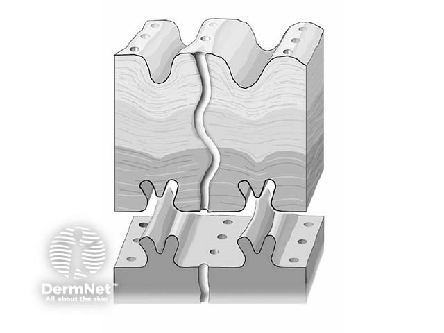
The epidermis has a complex structure designed to protect from the environment. It has an undulating surface with cross-crossing ridges and valleys, with invaginations due to follicles and sweat duct ostia. Epidermis is thickest on palms and soles, and thinnest on eyelid and scrotum.

Friction ridges
Ridges are particularly well developed on the fingers and toes where they are known as friction ridges with characteristic patterns commonly referred to as fingerprints. The science of ridgeology has been well developed for forensic purposes.

Friction ridges

Proliferation
Keratinocytes make up 95% of the skin surface and are normally renewed every 15 to 30 days. The speed of renewal is greater if the epidermis is injured and in certain skin diseases (particularly psoriasis). Keratinocytes are created in the basal layer and gradually move towards the surface, flattening out and becoming more differentiated towards the anuclear horny cell of the stratum corneum.

The appearance and structure of normal skin varies according to the site of origin of the tissue and the age, sex and ethnicity of the subject.

Epidermal cells

Basal cell layer
Keratinocytes
  • Columnar cells derived from ectoderm.
  • Stain with haematoxylin (i.e. pink on H&E sections).
  • Produce protein (keratin) and lipids.
  • Produce inflammatory cytokines including interleukin-1.
  • Express adhesion molecules.
  • Attached to surrounding cells by desmosomes.
  • Dividing cells with a roughly 19-day cycle. Daughter cells move to surface to form stratum corneum (28 to 60-days).
  • On haematoxylin and eosin (H&E) routine sections keratinocytes appear pink (taking up eosin dye) with blue nuclei (haematoxylin).
Melanocytes
  • Dendritic cells with clear cytoplasm and small dark-staining nuclei.
  • Derived from neural crest.
  • Ratio to basal keratinocytes is 1:10.
  • Produce melanin in melanosomes.
  • Dendritic processes allow transfer of melanin to adjacent keratinocytes by pinocytosis (tips of dendrites pinched off and engulfed).
  • Similar number of melanocytes in all races but melanogenesis is variable.
  • White skin: melanin mainly in basal layer. Blacks: melanin throughout epidermis. Tanning: melanin shifts into keratinocytes, production increased.
Merkel cells
  • Touch (sensory-mechanical) receptors.
  • Scarce small round cells found only using electron microscopy.
  • Most numerous in palms and soles.
  • Probably neural crest derived, possibly from keratinocytes.
  • Attached to keratinocytes by desmosomes.
Squamous cell layer (stratum spinosum, prickle cell layer)
Keratinocytes
  • 5-12 layers of polygonal cells that become flatter near surface.
  • Daughter cells produced by basal cells progress outwards.
  • Communicate with surrounding keratinocytes, melanocytes and Langerhans cells.
Langerhans cells
  • Dendritic immune cells with clear cytoplasm on haematoxylin-eosin (H&E) stained sections; identified by special stains (such as gold).
  • Characteristic “tennis racquet” granules on electron microscopy.
  • Derived from bone marrow.
  • Antigen-presenting cell: collects contact antigens and presents them to sensitised T lymphocytes.
  • Circulate via dermal lymphatics to regional lymph nodes.
  • Surface receptors for C3, Fc portion of IgG; express Ia antigens on surface.
Desmosomes
  • Attached to cell membrane opposite similar complex on adjacent cell.
  • Tonofilaments connect the keratinocyte cytoplasm with the desmosome.
  • In the intercellular space there is a lattice-work transmembrane linker.
  • Desmosomes make and break as keratinocytes move from basal layer to surface.
Inflammatory cells
  • Migrating neutrophils, eosinophils, lymphocytes, erythrocytes can be present in epidermis transiently in diseased states.
Granular cell layer (stratum granulosum)
Keratinocytes
  • 3- 5 layers of flattened cells filled with irregular dark keratohyaline granules.
Stratum lucidum
Keratinocytes
  • Thin homogeneous eosinophilic area (no organelles visible).
  • Cytoplasm contains keratin filaments.
  • Most obvious in areas of friction e.g. palms and soles.
Horny cell layer (stratum corneum)
Keratinocytes
  • Vertical stacks of flat cells move outwards over 14 days.
  • At base, adherent cell membranes.
  • At top, cells loosen and fall off.
  • Readily absorb and lose water.

Epidermal appendages

Structure Description
Eccrine glands
  • Sweat glands produce hypotonic solution of water, sodium chloride, urea, ammonia and uric acid.
  • Abundant, except vermilion of lips, labia minora, glans penis, prepuce.
  • Most dense on palms, soles, axillae and forehead.
  • Sweat on palms and soles enhances grip.
  • Secretory coil deep in dermis, duct opens directly onto skin surface.
  • Duct composed of two layers of small cuboidal cells.
  • Immune function: secrete IgA.
  • Hypothalamic control via specific nerve fibres results in increased production with heat, emotional stress and spicy foods.
  • Develop tolerance to high ambient temperatures by increased sweat production and increasing hypotonicity.
Apocrine glands
  • Scent glands that become active after puberty.
  • Mainly found in axillae and perianal regions.
  • Primary secretion is thick and odourless; smell derives from bacterial colonisation.
  • Modified as Moll’s glands (eyelids), ceruminous glands in ear canal, mammary glands,
  • Open into pilosebaceous follicle.
Pilosebaceous structures
  • Terminal hair on scalp; vellus hair on body surface (short, thin, light coloured).
  • None on palms and soles.
  • Root sheath buds downward from epithelium at a consistent angle, depending on site.
  • Growth phase (anagen) with pointed tip lasts several years; short involutional phase (catagen); resting phase for several months, with clubbed or bulbous tip (telogen).
  • Hair cortex is produced at a rapid metabolic rate from medulla (loose cuboidal cells) within hair bulb. Cortex contains densely packed keratin with extra sulphur and cystine.
  • Cortex surrounded by cuticle: a single layer of shingle-like cells.
  • Hair colour depends on amount of melanin in cortex during anagen: dark hair has more eumelanin melanosomes; fair hair fewer with more lamellated phaeomelanin; red hair has erythromelanin; grey/white hair very few melanocytes.
Anatomy and physiology of hair
Sebaceous glands
  • Most concentrated on scalp and face where circulating androgens induce increased secretion at puberty.
  • Several lobules lead into common excretory duct, mostly opening into outer portion of hair follicle. Opens directly onto skin surface on labia, prepuce, nipple and areola.
  • Modified in Montgomery’s tubercles of areolae secreting into lactiferous duct.
  • Produce sebum: triglycerides, phosopholipids, esterified cholesterol.
Nails
  • Nail matrix produces cornified cells of nail plate; outer cells formed by proximal matrix, innermost by distal matrix (lunula).
  • Fingernails grow 0.1mm per day; toenails 0.03mm per day.
  • Cuticle protects matrix from environment.
  • Nails protect and scratch.
Normal nail

Activity

Download and label the components shown on the Diagram: Normal Epidermis Empty – PDF file

Check the answers: Normal Epidermis – PDF file

Take the Structure of the epidermis quiz


Related information

References

On DermNet NZ

Information for patients

Other websites

Books about skin diseases

Sign up to the newsletter