Main menu
Common skin conditions

NEWS
Join DermNet PRO
Read more
Quick links

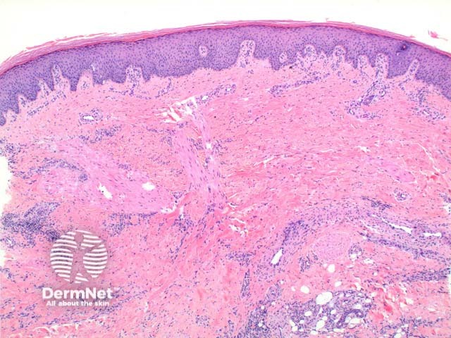
Figure 2
Keywords: Necrobiosis lipoidica, Histopathology-image, Pathology
Scanning power view of necrobiosis lipoidica demonstrates a layered inflammatory process and alternating zones of necrobiosis involving the full thickness of the dermis (Figure 1). The changes tend to become more pronounced deeper in the dermis and may extend into the septal panniculus (Figures 2 and 3). The areas of necrobiosis are poorly defined and run into each other with broad foci of inflammatory infiltrate intervening (Figure 4). This may form a stacked ‘lasagne’ type appearance. A variable histiocytic infiltrate with multinucleated giant cells surrounds these areas. The accompanying inflammatory infiltrate is predominantly lymphocytic with plasma cells and occasional eosinophils (Figure 5). As lesions age an increasing degree of dermal fibrosis is seen.
© DermNet
You can use or share this image if you comply with our image licence. Please provide a link back to this page.
For a high resolution, unwatermarked copy contact us here. Fees apply.
Source: dermnetnz.org