

Epidermolytic hyperkeratosis pathology — extra information
Introduction
Epidermolytic hyperkeratosis is a histological pattern seen in isolation or as an incidental finding in a number of dermatological conditions.
Histology of epidermolytic hyperkeratosis
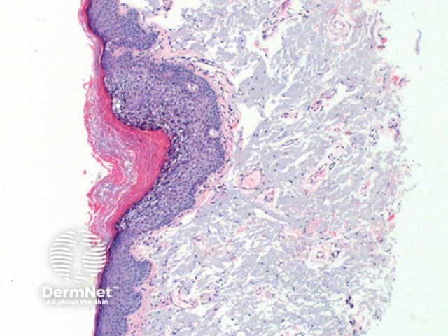
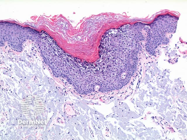
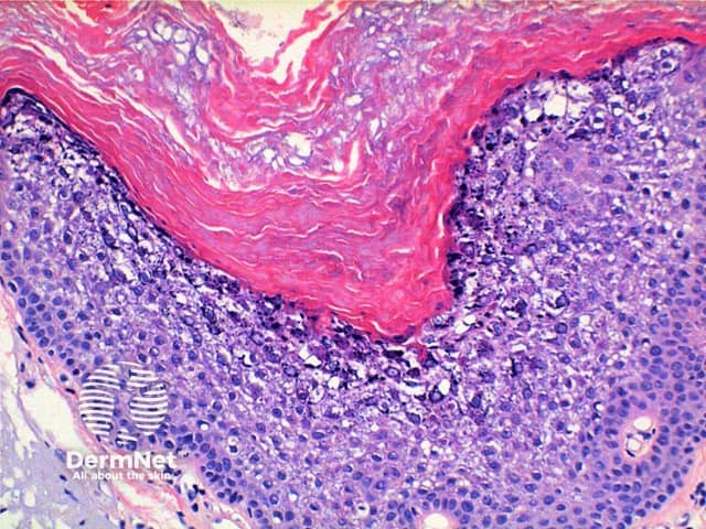
Low power view of histology of epidermolytic hyperkeratosis demonstrates hyperkeratosis and epidermal hyperplasia of varying degrees (Figure 1). The diagnostic features include a characteristic vacuolar degeneration with hypergranulosis of the stratum granulosum and stratum spinosum (Figures 2 and 3).
 pathology
" data-source="dermnetnz.org" data-id="12232" data-url="12232-epidermolytic-hyperkeratosis-pathology">
Histological variants of epidermolytic hyperkeratosis
Epidermolytic acanthoma: When the changes of epidermolytic hyperkeratosis are seen forming a solitary lesion. Rarely multiple discrete lesions may be seen in disseminated epidermolytic acanthoma.
Epidermolytic ichthyosis: epidermolytic hyperkeratosis may be seen within biopsies of this generalised congenital condition.
Incidental: Epidermolytic hyperkeratosis may be seen in normal skin adjacent to any skin lesion or dermatosis.
Epidermolytic leukoplakia: is the term used for epidermolytic features arising on a mucosal surface (which is nonkeratinised).
Epidermal naevus variant: epidermolytic hyperkeratosis may be seen within some linear and systematised epidermal naevi.
References
- Skin Pathology (3rd edition, 2002). Weedon D
- Pathology of the Skin (3rd edition, 2005). McKee PH, J. Calonje JE, Granter SR
On DermNet
Other websites
- Epidermolytic Hyperkeratosis (Bullous Congenital Ichthyosiform Erythroderma) — eMedicine Dermatology