Folliculosebaceous cystic hamartoma represents a cutaneous biphasic hamartoma. It is composed of epithelial and mesenchymal components in varying proportions, typically occurring in adults in the head and neck area. Several pathogenetic mechanisms underlying this adnexal neoplasm have been proposed. However, there remains much controversy as to whether it represents a distinct entity or part of an evolutionary spectrum of trichofolliculomas (TF). Since the introduction of this entity in 1991, there have been some case reports documenting a range of mesenchymal changes seen in FCH as well as uncommon and unusual associations.
Folliculosebaceous cystic hamartoma is a cutaneous hamartoma of epithelial and mesenchymal elements and is primarily centred on the dermis. The epithelial components of folliculosebaceous cystic hamartoma comprise distorted folliculosebaceous cystic proliferations while mesenchymal components commonly include fibroplasia, vascular and adipose tissue proliferations.
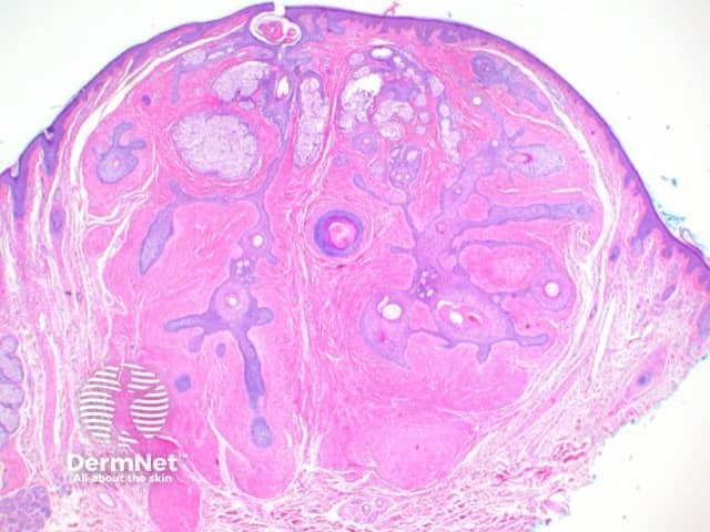
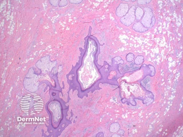
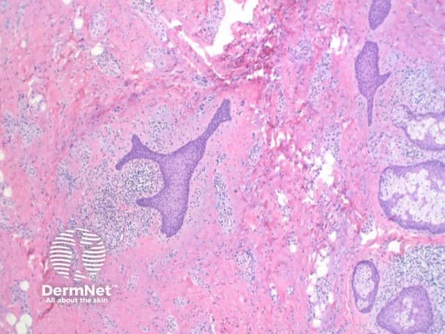
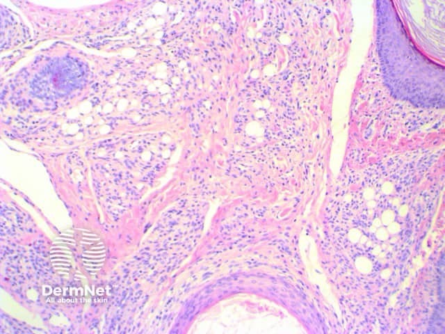
Scanning view shows dilated follicular structure or structures, with radiating mature sebaceous lobules (figures 1, 2). Surrounding the distorted hair follicles there may be thin anastomosing epithelial strands (figure 3). The stroma surrounding the epithelial units is frequently composed of dense collagenous tissue (figure 1-3) and can show prominent clefting to the surrounding adjacent uninvolved dermis (figure 1). Additional stromal changes can include increased vascularity or pockets of adipocytes consistent with increased stromal fat (figure 4).
Trichofolliculoma: Attempts to distinguish sebaceous trichofolliculoma from folliculosebaceous cystic hamartoma on clinical and histologic grounds have been made. It has been suggested the latter lesion clinically presents as a papulonodular lesion without hairs. Histologically, FSH demonstrates more prominent stromal mesenchymal abnormalities.
In contrast, sebaceous trichofolliculoma clinically presents as a depressed lesion with an eruption of terminal or vellus hairs. Histologically, there is an almost constant connection of the dilated infundibular structure with the epidermis. However, the features described above are not absolute. There have been clinical descriptions of folliculosebaceous cystic hamartoma associated with hair, and hair shafts have also been identified within cystic structures in folliculosebaceous cystic hamartoma. Similarly, the finding of a direct connection to the epidermis by the cystic structure is not exclusively found in sebaceous trichofolliculoma and have been described in cases of folliculosebaceous cystic hamartoma.

