Glomangiomas (also called glomuvenous malformation) differ clinically from glomus tumors in that they occur in childhood and adolescence, are usually asymptomatic, do not have a predilection for the subungal region, and often are multifocal. They can vary in colour from pink-to-blue and often become darker with age; they may be plaque-like or nodular. Multiple glomangiomas are rare and comprise about 10 percent of all glomus tumours.
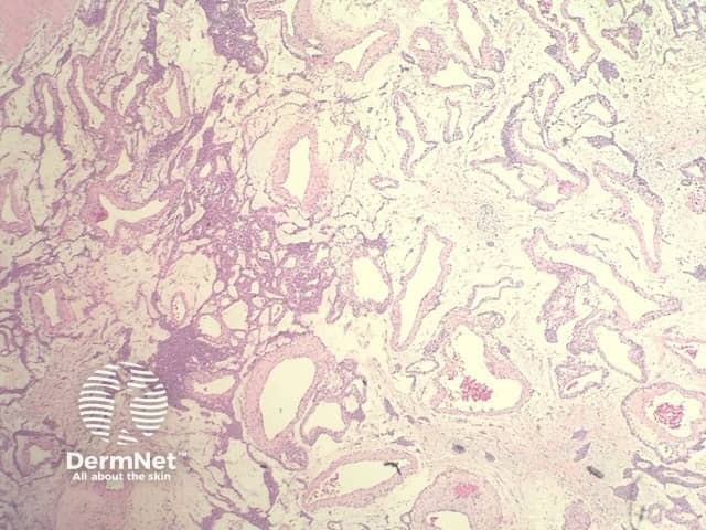
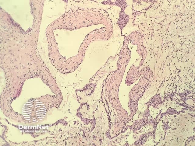
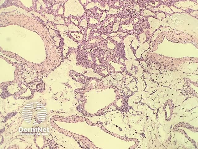
In glomangioma, the histopathology shows dilated venous channels that resemble venous malformations (figures 1, 2). Unlike venous malformations, they demonstrate single to multiple rows of surrounding cuboidal glomus cells (figures 3,4).
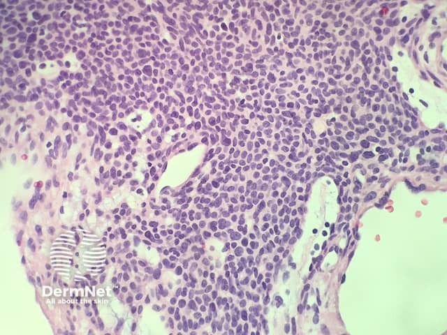
The glomus cells stain positively for vimentin and α-smooth-muscle actin but are negative for desmin, von Willibrand factor, and S-100.
Other diagnoses to be considered include:

