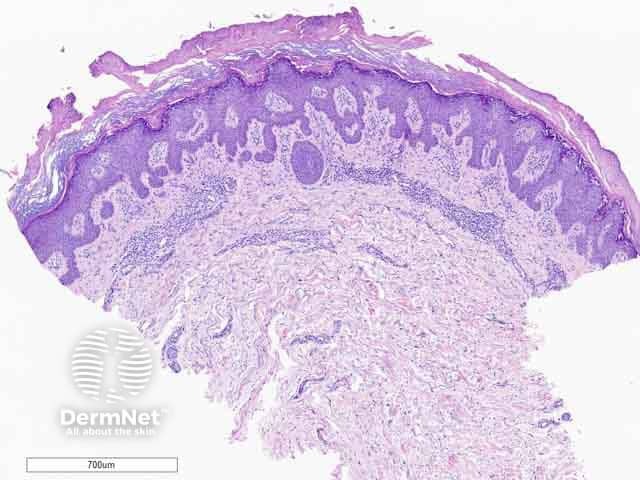
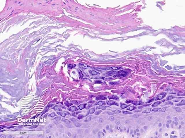
Hypergranulotic dyscornification is a histological reaction pattern seen in benign cutaneous keratoses.
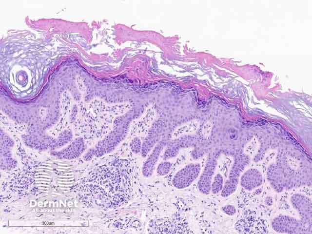
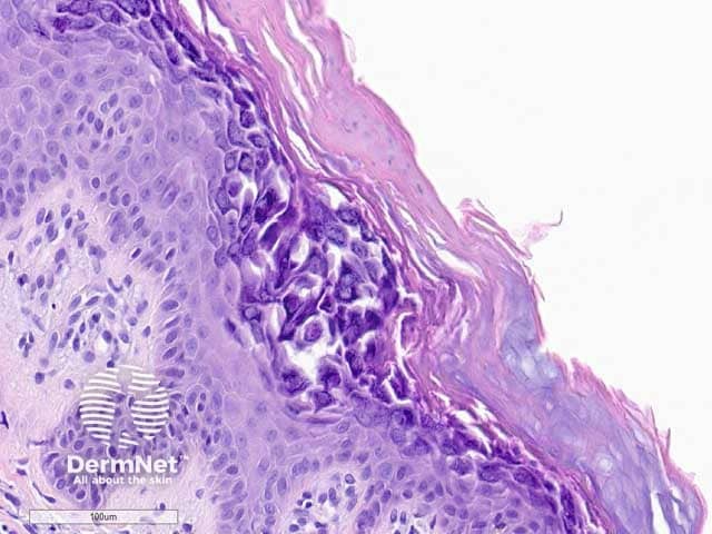
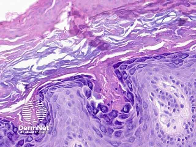
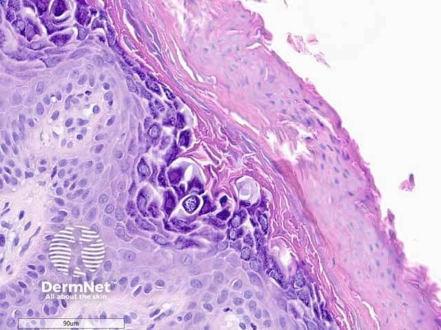
Hypergranulotic dyscornification is reminiscent of verruca vulgaris. The lesion may be exoendophytic (figure 1). There are finger-like projections of epidermal hyperplasia and hypergranulosis with clustered keratohyalin granules. The entire lesion shows compact orthokeratosis underneath a laminated and basket-weave stratum corneum (figure 2). The key feature is the corneocytes in the stratum corneum which appear rounded, glassy, and eosinophilic (figures 3–6). Parakeratosis is also usually present. There is a variable underlying inflammatory lymphocytic infiltrate in the upper dermis.