

Hypergranulotic dyscornification pathology — extra information
Synonyms:
Categories:
ICD-10:
ICD-11:
SNOMED CT:
ADVERTISEMENT
What is hypergranulotic dyscornification?
Hypergranulotic dyscornification is a histological reaction pattern seen in benign cutaneous keratoses.
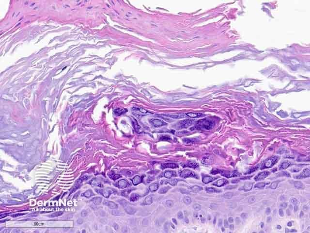
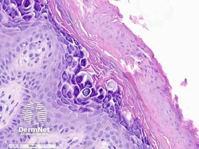
Histology of hypergranulotic dyscornification
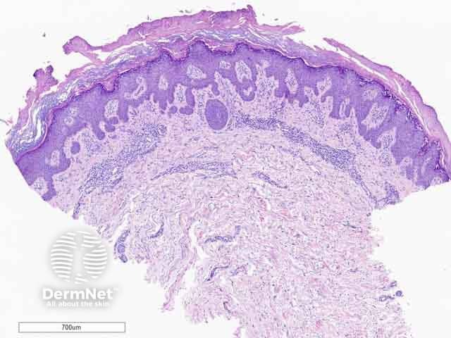
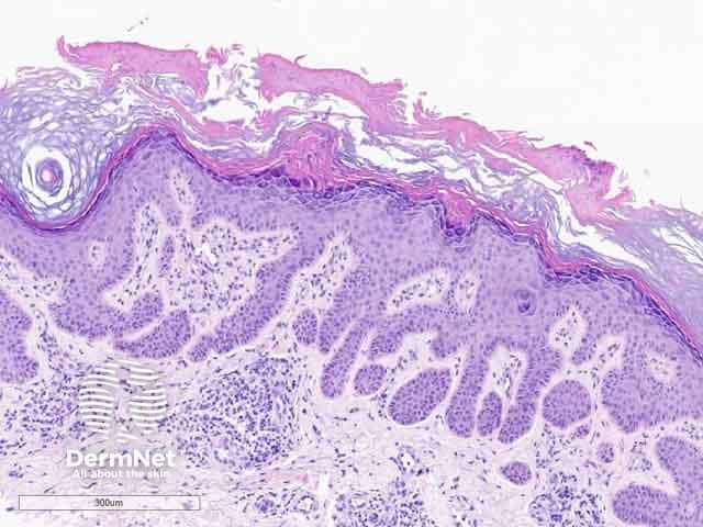
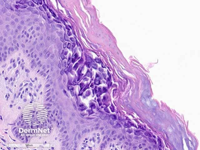
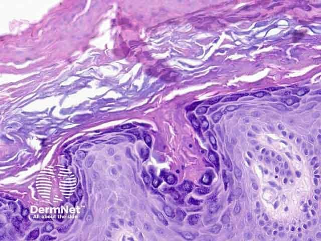
Hypergranulotic dyscornification is reminiscent of verruca vulgaris. The lesion may be exoendophytic (figure 1). There are finger-like projections of epidermal hyperplasia and hypergranulosis with clustered keratohyalin granules. The entire lesion shows compact orthokeratosis underneath a laminated and basket-weave stratum corneum (figure 2). The key feature is the corneocytes in the stratum corneum which appear rounded, glassy, and eosinophilic (figures 3–6). Parakeratosis is also usually present. There is a variable underlying inflammatory lymphocytic infiltrate in the upper dermis.
Differential diagnosis of hypergranulotic dyscornification pathology
- Epidermolytic hyperkeratosis. The keratohyalin granules are not as thick and clumped, but instead small and dotted in epidermolytic hyperkeratosis. The eosinophilic corneocytes seen in hypergranulocytic dyscornfication are also not present. There is usually reticular degeneration present in epidermolytic hyperkeratosis.
- Verruca vulgaris. Koilocytes are present in verruca vulgaris and not seen in hypergranulotic dyscornification.
- Seborrhoeic keratosis. Horn pseudocysts will usually be present in seborrhoeic keratosis and absent in hypergranulotic dyscornification.
Bibliography
- Gesheva A, Pitch M, Rosamilia L, Hossler E. Keratotic papule on the abdomen. Cutis. 2020;105(5):222–31. Journal
- Patterson JW. Weedon’s Skin Pathology, 5th edn. Elsevier, 2021. p336.
- Reichel M. Hypergranulotic dyscornification: a distinctive histologic pattern of maturation of epidermal epithelium present in solitary keratoses. Am J Dermatopathol. 1999;21(1):21–4. doi:10.1097/00000372-199902000-00004 PubMed
- Roy SF, Ko CJ, Moeckel GW, Mcniff JM. Hypergranulotic dyscornification: 30 cases of a striking epithelial reaction pattern. J Cutan Pathol. 2019;46(10):742–7. doi:10.1111/cup.13522. PubMed
On DermNet
Books about skin diseases
ADVERTISEMENT
Other recommended articles
ADVERTISEMENT