

Infective panniculitis — extra information
Introduction
Clinical features
Diagnosis
Bacterial causes
Fungal panniculitis
Viral panniculitis
What is infective panniculitis?
Panniculitis of the skin is inflammation of the subcutaneous fat. It has many causes. An infective cause is more likely in those with immunodeficiency (a weakened immune system).
Immunodeficiency may be due to disease, such as infection with human immunodeficiency virus (HIV), or drug-induced immunosuppression (eg, prescribed after organ transplantation). Immunodeficiency may lead to infection with unusual organisms in unusual sites, including the subcutaneous fat.
How does infective panniculitis present?
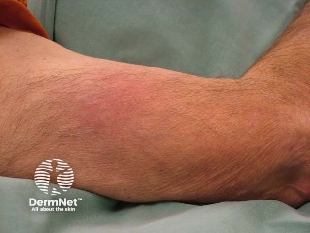
Infective panniculitis, in general, has no specific features. The clinical presentation depends on:
- The type of organism
- Its virulence (how harmful it is)
- The route of inoculation
- The immune state of the individual.
How is the diagnosis of infective panniculitis made?
Whilst history is important, the diagnosis usually requires microbiological and histological confirmation. On biopsy, most infectious panniculitis causes a lobular pattern of inflammation. A lobular pattern is nonspecific although there are sometimes clues that an organism is responsible. Cultures and special stains are required to distinguish these.
Bacterial causes of panniculitis
Common bacteria that cause panniculitis include:
- Streptococcus pyogenes
- Staphylococcus aureus
- Pseudomonas spp
- Klebsiella spp
- Nocardia spp
- Brucella spp
Bacterial panniculitis can appear in immunocompetent as well as immunosuppressed individuals. It can develop as a result of direct inoculation or from seeding from systemic infection.
The classical histopathological appearance is a lobular or mixed lobular/septal suppurative panniculitis with heavy infiltrate of neutrophils.
The choice of antibiotic treatment depends on the specific bacteria identifed. If the microorganism is unknown, a broad-spectrum antibiotic such as amoxicillin-clavulanic acid is often chosen.
Mycobacterial panniculitis
Mycobacteria have a wide range of skin manifestations and panniculitis is uncommon. Most mycobacterial panniculitis is due to atypical mycobacteria including:
- Mycobacterium chelonae
- Mycobacterium fortuitum
- Mycobacterium abscessus
- Mycobacterium avium-intracellulare complex
- Mycobacterium marinum
- Mycobacterium leprae (leprosy, rare)
- Mycobacterium ulcerans
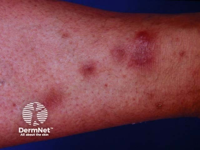
Mycobacterial panniculitis is most frequently seen in immunocompromised individuals spreading through the bloodstream to present as widespread skin lesions. Trauma is the usual means of inoculation in individuals with a normal immune system and results in a single lesion.
Histology demonstrates lobular panniculitis, sometimes with granuloma formation. Zeihl-Neelson, auramine-rhodaine or Fite-Faraco stains are specialised mycobacterial stains that help highlight the organism. Cultures are more sensitive and allow for accurate identification of the mycobacterial species but can take many weeks. DNA probes and DNA PCR are quick ways of species identification.
Treatment regimes are specific to the species of mycobacteria. Treatment is usually continued for 6–12 months or at least 6–8 weeks after clinical resolution.
Treatment of cutaneous non-tuberculous mycobacterial infection |
||
|---|---|---|
Micro-organism |
First Line |
Other treatment options |
M. chelonae |
Clarithromycin + ciprofloxacin/doxycycline |
Surgical debridement Dual antimicrobial therapy |
M. fortuitum |
Amikacin + ciprofloxacin/doxycycline |
Surgical debridement Dual antimicrobial therapy |
M. abscessus |
Clarithromycin + amikacin/cefoxitin |
Surgical debridement/excision |
M. marinum |
Ethambutol + rifampicin or doxycycline |
Surgical debridement |
M. avium-intracellulare |
Ethambutol + clarithromycin + rifampicin |
Surgical excision |
Fungal panniculitis
Fungal panniculitis can be separated into disseminated disease or classical subcutaneous mycosis.
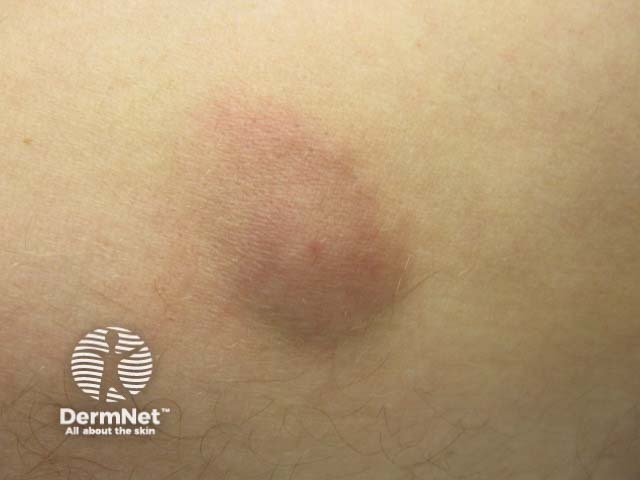
- A disseminated fungal infection affects very unwell immunocompromised individuals, who present with multiple inflamed subcutaneous nodules.
- Classical subcutaneous mycosis develops in healthy individuals as a single nodule on the lower leg and then becomes an ulcer or abscess that does not respond to antibiotics.
Biopsy, fungal culture and specific histology stains (silver-impregnated procedure and periodic acid-Schiff) are required to make the diagnosis.
Treatment of fungal panniculitis depends on the identification of the organism.
- Disseminated fungal infections carry high mortality and require a long duration of antifungal treatment unless the immune system is reconstituted.
- Classical subcutaneous mycosis is extremely difficult to cure and a long duration of antifungal treatment is required often preceded by complete surgical excision.
Features of deep fungal infections |
||
|---|---|---|
Disseminated fungal disease |
Classical subcutaneous mycosis |
|
Causative organisms |
Candida spp, Aspergillus spp, Fusarium spp, Histoplasma capsulatum |
[Sporotrichosis] (/topics/sporotrichosis) (Sporothrix schenckii), eumycetoma (Madurella mycetomatis), chromoblastomycosis (Phialophora verrucosa, Fonsecaea pedrosoi and F. compacta), zygomycosis (Entomophthorales) |
Patient characteristics |
Immunosuppressed individuals |
A healthy individual directly inoculated by soil, plant, or wood |
Clinical manifestations |
Multiple inflammatory subcutaneous lesions Individual very unwell |
Single slowly growing subcutaneous nodule Can discharge pus and invade deep tissues |
Histology |
Lobular panniculitis without vasculitis |
Lobular panniculitis without vasculitis Occasionally, suppurative granuloma |
Management |
Therapy may include itraconazole or amphotericin B (for months) Surgical excision may be needed for some lesions |
Surgical excision Itraconazole (months) |
Viral panniculitis
The medical literature about viral panniculitis is limited to a few case reports. Cytomegalovirus (CMV) has been reported to cause panniculitis in an immunocompromised patient.
Bibliography
- Delgado-Jimenez Y, Fraga J, García-Díez A. Infective panniculitis. Dermatol Clin. 2008;26(4):471-vi. doi:10.1016/j.det.2008.05.005 PubMed
- Morrison LK, Rapini R, Willison CB, Tyring S. Infection and panniculitis. Dermatol Ther. 2010;23(4):328–40. doi:10.1111/j.1529-8019.2010.01333.x PubMed
- Torrelo A, Hernández A. Panniculitis in children. Dermatol Clin. 2008;26(4):491-vii. doi:10.1016/j.det.2008.05.010. PubMed
On DermNet