

Maculopapular cutaneous mastocytosis — extra information
Introduction
In children
In adults
Precautions
Investigations
Treatment
What is maculopapular cutaneous mastocytosis?
Maculopapular cutaneous mastocytosis, also called urticaria pigmentosa, is due to abnormal collections of mast cells in the skin causing brown patches and freckles. It is the most common type of cutaneous mastocytosis.
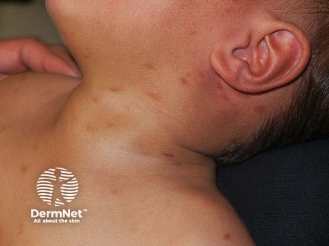
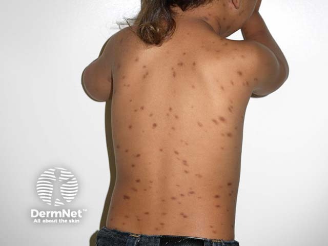
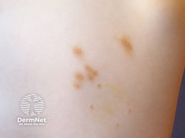
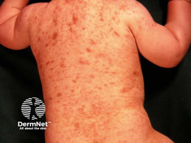
Maculopapular cutaneous mastocytosis in children
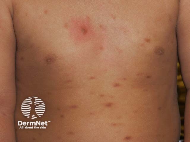
Maculopapular cutaneous mastocytosis most often affects infants, with the first patches appearing at a few months of age. They patches are often confused with moles or with insect bites at first, but the lesions persist and gradually increase in number for several months or years. They can appear on any part of the body including the scalp, face, trunk and limbs.
In young children, it is common for the mastocytosis patches to blister when rubbed. If many patches are rubbed or activate spontaneously at the same time, the infant may become irritable, but is uncommon for severe symptoms to arise.
Over time, the mastocytosis becomes less itchy and eventually the patches fade away. By the teenage years, most patches will have gone.
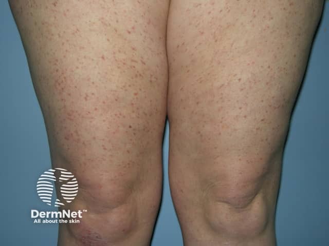
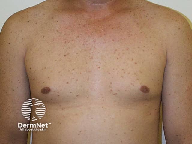
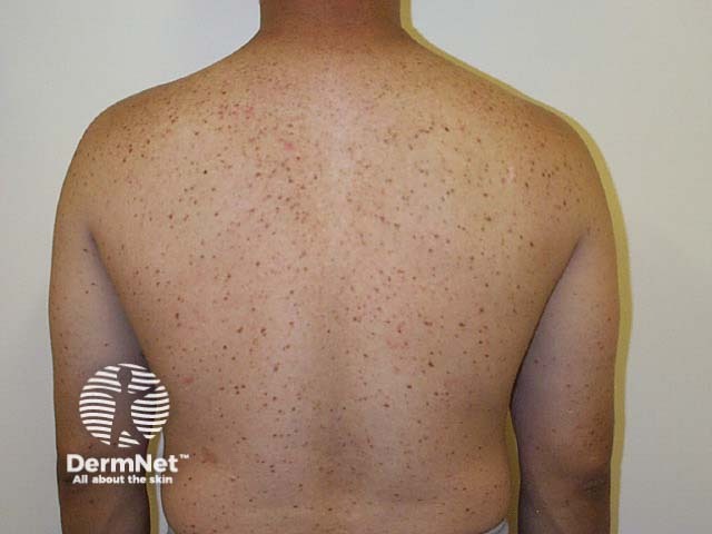
Maculopapular cutaneous mastocytosis in adults
Maculopapular cutaneous mastocytosis can sometimes develop for the first time in an adult. Just a few lesions may appear, or many. The lesions can be unsightly and itchy. Unfortunately, in adults, urticaria pigmentosa tends to persist long-term. Sometimes patients can have systemic symptoms (such as flushing, itching, low blood pressure, anaphylactic shock, diarrhoea and bleeding from the gastrointestinal tract), which suggest the presence of systemic mastocytosis.
Localised cutaneous mastocytosis has rarely been reported to arise in an area of radiation treatment for breast cancer for unknown reasons.

What precautions should I take?
Exercise or heat can aggravate symptoms of cutaneous mastocytosis. A severe reaction can result in flushing and faintness.
Certain medications can cause mast cell degranulation and should be avoided if there is extensive urticaria pigmentosa. These include:
- Aspirin (salicylates) and other nonsteroidal anti-inflammatory drugs
- Codeine and morphine (narcotics)
- Alcohol
- Anticholinergics.
People with urticaria pigmentosa may get a severe allergic reaction from bee or wasp stings, hence they may need to carry an injectable adrenaline (epinephrine) kit.
What investigations should be done?
The appearance of maculopapular cutaneous mastocytosis is generally so characteristic that no specific tests are necessary. However, occasionally a skin biopsy is needed to confirm the diagnosis (see maculopapular cutaneous mastocytosis pathology). If there are any symptoms suggesting internal involvement (systemic mastocytosis), refer to our page on mastocytosis for further information. Tests may include:
- Blood count
- Serum tryptase
- DEXA bone scan
- Bone marrow examination
What is the treatment for maculopapular cutaneous mastocytosis?
Maculopapular cutaneous mastocytosis is not serious, and does not require any treatment in most cases (unless there is also systemic involvement). The following can be helpful.
- Oral antihistamines
-
Topical steroids
Potent steroid creams applied for several months under occlusion can reduce itching and unsightliness, but the patches tend to recur within a few months. Topical steroids are only suitable for limited areas. - Topical calcineurin inhibitors, such as pimecrolimus cream
-
Phototherapy
This form of ultraviolet radiation is the most effective treatment for adults with maculopapular cutaneous mastocytosis. Two or three treatments each week are required for several months. Narrowband UVB or (if available) photochemotherapy (PUVA) lessens the itch and improves the appearance. The cutaneous mastocytosis is likely to recur within six to twelve months.
References
- Azaña JM, Torrelo A, Matito A. Update on mastocytosis (Part 2): categories, prognosis, and treatment. Actas Dermosifiliogr. 2016 Jan-Feb;107(1):15–22. doi:10.1016/j.ad.2015.09.009. English, Spanish. PubMed PMID: 26525106. PubMed.
- Castells M, Metcalfe DD, Escribano L. Diagnosis and treatment of cutaneous mastocytosis in children: practical recommendations. Am J Clin Dermatol. 2011 Aug 1;12(4):259-70. doi: 10.2165/11588890-000000000-00000. Review. PubMed PMID: 21668033; PubMed Central PMCID: PMC4126834.PubMed Central
- Hartmann K, Escribano L, Grattan C, et al. Cutaneous manifestations in patients with mastocytosis: Consensus report of the European Competence Network on Mastocytosis; the American Academy of Allergy, Asthma & Immunology; and the European Academy of Allergology and Clinical Immunology. J Allergy Clin Immunol. 2016 Jan;137(1):35–45. doi: 10.1016/j.jaci.2015.08.034. Review. PubMed PMID: 26476479. PubMed.
- Mashiah J, Harel A, Bodemer C, Hadj-Rabia S, Goldberg I, Sprecher E, Kutz A. Topical pimecrolimus for paediatric cutaneous mastocytosis. Clin Exp Dermatol. 2018 Feb 20. doi: 10.1111/ced.13391. PubMed PMID: 29460435. PubMed.
- Sandru F, Petca RC, Costescu M, et al. Cutaneous mastocytosis in childhood - update from the literature. J Clin Med. 2021;10(7):1474. doi:10.3390/jcm10071474 Journal
On DermNet
- Maculopapular cutaneous mastocytosis pathology
- Mastocytosis
- Mastocytoma
- Urticaria
- Sensitive skin
- Skin manifestations of haematological disease
- Exercise-induced anaphylaxis
- Blisters and pustules in neonates
Other websites
- Australasian Mastocytosis Society
- The Mastocytosis Society, Inc.
- Mastocytosis — Medscape Reference
- Urticaria Pigmentosa — British Association of Dermatologists