

Necrolytic acral erythema — extra information
Introduction Demographics Causes Clinical features Complications Diagnosis Differential diagnoses Treatment Outcome
What is necrolytic acral erythema?
Necrolytic acral erythema is a clinically distinct entity affecting the skin of the toes and tops of feet, seen predominantly in patients with hepatitis C.
Who gets necrolytic acral erythema?
Necrolytic acral erythema was first described in hepatitis C-positive patients. A study in the US suggested it develops in 1–2% of patients with chronic hepatitis C, and most studies report this to be the commonest association for this skin sign [see Viral hepatitis].
A small number of affected patients are seronegative for hepatitis C. The next most common association reported has been zinc deficiency occurring with various other disorders including coeliac disease and Crohn disease.
All reported cases have been in skin of colour including Asians and Africans, with an equal sex incidence, and a mean onset of 40–45 years of age (range 11–78 years).
What causes necrolytic acral erythema?
The aetiopathogenesis of necrolytic acral erythema appears to be multifactorial and may involve genetic factors, nutritional deficiency (specifically zinc), and liver dysfunction resulting in low blood levels of albumin, glucagon, and amino acids.
Although Japan has a very high rate of chronic hepatitis C infection, no cases have been reported from Japan. This may reflect the predominant hepatitis C genotypes in that population or predisposing genetic factors.
Zinc is an essential cofactor for replication of the hepatitis C viral genome.
Viral load and immunosuppression do not appear to be risk factors.
What are the clinical features of necrolytic acral erythema?
Necrolytic acral erythema can be the first sign of hepatitis C infection or zinc deficiency, presenting as a symmetrical acral rash.
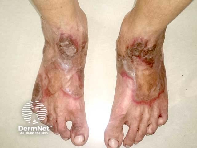
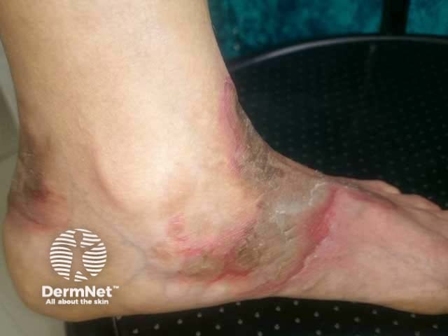
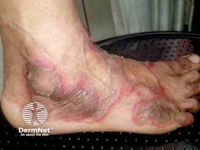
Necrolytic acral erythema typically affects the tops of the toes and feet. Sometimes the skin lesions occur on the ankles, knees, and legs, and rarely the hands, elbows, genitalia, and buttocks. Palms, soles, and nails are almost always spared.
The skin lesions are usually painful or sometimes itchy or with a burning sensation.
Acute necrolytic acral erythema
The acute skin lesions are red with a margin of erosions or flaccid blisters.
Chronic necrolytic acral erythema
- Initial phase: papules and plaques
- Scaly
- Dusky or eroded centre.
- Well-developed phase: plaques
- Papules and plaques coalesce
- Large, well-demarcated
- Hyperpigmented, thick with adherent scale
- Occasional pustules.
- Late phase: plaques
- Thin, circumscribed, psoriasiform
- Hyperpigmented
- Well-defined dark red rim.
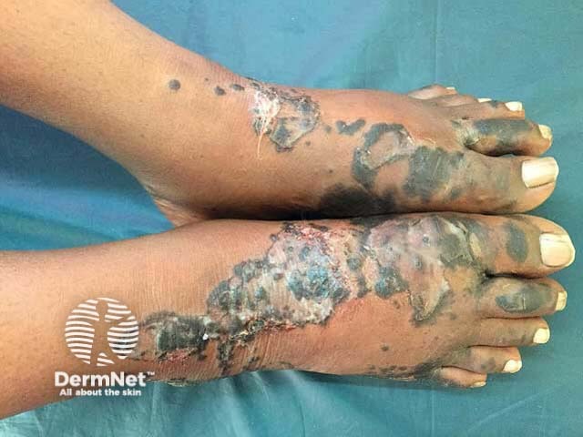
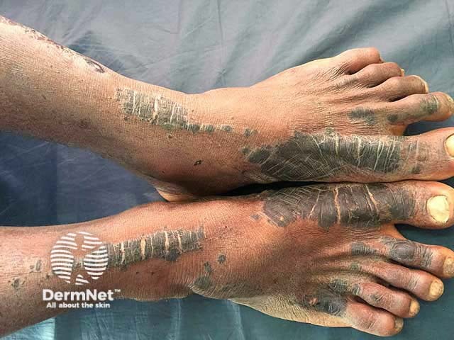
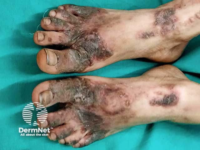
Images reproduced with permission from the authors: Inamadar AC, Shivanna R, Ankad BS. Necrolytic acral erythema: current insights. Clin Cosmet Investig Dermatol. 2020;13:275–81.
Dermoscopy of necrolytic acral erythema
- Brown-white background
- Dull white scale
- Central white and yellow globules, brown globules at the periphery
- Scattered irregular red dots in the centre
What are the complications of necrolytic acral erythema?
- Lichenification
- Fissuring
- Consequences of missing the diagnosis of hepatitis C infection
How is necrolytic acral erythema diagnosed?
Necrolytic acral erythema should be suspected on the skin signs in an appropriate clinical setting.
Skin biopsy shows the same histological features as necrolytic migratory erythema: hyperkeratosis with parakeratosis, epidermal spongiosis and focal necrosis, mixed superficial dermal inflammatory infiltrate.
Blood tests should include:
- Liver function tests
- Hepatitis C serology
- Serum zinc
- Other tests as clinically indicated — may include hepatitis B and HIV serology due to the similar mode of transmission.
What is the differential diagnosis for necrolytic acral erythema?
- Psoriasis
- Necrolytic migratory erythema
- Other nutrition-related skin conditions: pellagra, acrodermatitis enteropathica-like rashes, biotin responsive dermatoses
What is the treatment for necrolytic acral erythema?
- Oral zinc supplements — 220 mg twice daily
- Treatment of associated disorders
- Hepatitis C
- Malabsorption
Topical treatments, such as topical steroids or calcineurin inhibitors (eg, pimecrolimus, tacrolimus), do not seem to be helpful.
What is the outcome for necrolytic acral erythema?
Necrolytic acral erythema follows a relapsing course of spontaneous remissions and recurrences. Skin changes rapidly settle with appropriate treatment, but can recur if treatment is stopped.
Bibliography
- el Darouti M, Abu el Ela M. Necrolytic acral erythema: a cutaneous marker of viral hepatitis C. Int J Dermatol. 1996;35(4):252–6. doi:10.1111/j.1365-4362.1996.tb02997.x. PubMed
- Das A, Kumar P, Gharami RC. Necrolytic acral erythema in the absence of hepatitis C virus infection. Indian J Dermatol. 2016;61(1):96–9. doi:10.4103/0019-5154.174047. Journal
- Davis S, Creditt A. Bilateral foot skin eruption in a hepatitis C patient. Clin Pract Cases Emerg Med. 2020;4(3):491–2. doi:10.5811/cpcem.2020.7.46490. PubMed Central
- Inamadar AC, Shivanna R, Ankad BS. Necrolytic acral erythema: current insights. Clin Cosmet Investig Dermatol. 2020;13:275–81. doi:10.2147/CCID.S189175. Journal
- Khader A, Sreekanth S, Manakkattu SH, John N. Necrolytic acral erythema as a manifestation of Crohn’s disease and celiac disease – a report of two cases. J Skin Sex Trans Dis. 2020;2:130–3. doi: 10.25259/JSSTD_17_2020. Journal
- Kumar R, Arora S, Ranjan E, Das N. Necrolytic acral erythema in seronegative hepatitis C patient with vitamin B12 deficiency. Indian Dermatol Online J. 2020;11(2):278–9. doi:10.4103/idoj.IDOJ_398_19. PubMed Central
- Raphael BA, Dorey-Stein ZL, Lott J, Amorosa V, Lo Re V 3rd, Kovarik C. Low prevalence of necrolytic acral erythema in patients with chronic hepatitis C virus infection. J Am Acad Dermatol. 2012;67(5):962–8. doi:10.1016/j.jaad.2011.11.963. PubMed Central
- Shah P, Ovits C, Brinster N, Lo Sicco K. Necrolytic acral erythema in a patient with sarcoidosis. JAAD Case Rep. 2020;6(11):1162–4. doi:10.1016/j.jdcr.2020.04.035. PubMed Central
- Srisuwanwattana P, Vachiramon V. Necrolytic acral erythema in seronegative hepatitis C. Case Rep Dermatol. 2017;9(1):69–73. doi:10.1159/000458406. Journal
- Xue R, Elbendary A, Wu T. Necrolytic acral erythema in a Chinese patient with hepatitis C and hepatitis B virus coinfection. An Bras Dermatol. 2019;94(4):446–8. doi:10.1590/abd1806-4841.20197257. PubMed Central
On DermNet
Other websites
- What is necrolytic acral erythema? — Medscape